Category: Chattanooga

  • How to tell if a mole is cancerous: the ABCDE rule explained

    How to tell if a mole is cancerous: the ABCDE rule explained

    Skin cancer is statistically the most common form of cancer in the United States. In fact, it is estimated that one in five Americans will develop skin cancer in their lifetime. While those numbers can be daunting, there is a powerful silver lining: skin cancer, including the most aggressive form, melanoma, has an exceptionally high cure rate when detected and treated in its earliest stages.

    At Chattanooga Skin and Cancer Clinic, our board-certified dermatologists and residency-trained specialists believe that the best defense is a proactive offense. Education is the first step in that defense. By understanding the ABCDE rule, you become an active participant in your own healthcare, capable of spotting changes that might otherwise go unnoticed.

    Understanding Melanoma: Why the ABCDEs Matter
    Before we dive into the specific rules, it is important to understand what we are looking for. Most benign (non-cancerous) moles are made of melanocytes (pigment-producing cells) that grow in a cluster but behave normally. Melanoma occurs when the DNA in those cells becomes damaged, usually due to UV radiation from the sun or tanning beds, causing them to grow uncontrollably and invade surrounding tissue.

    Because melanoma has the potential to spread (metastasize) to other organs, time is of the essence. The ABCDE rule was developed by dermatologists to provide the public with a clear, easy-to-remember framework for identifying these cellular malfunctions before they become life-threatening.

    The ABCDE Rule: A Deep Dive into the Red Flags

    A is for Asymmetry
    In the world of dermatology, symmetry is a sign of stability. If you were to draw an imaginary line down the center of a healthy mole, the two halves should be mirror images of each other. This indicates that the cells are growing at a uniform, controlled rate.
    The Red Flag: If you find a mole where the two halves do not match, it is considered asymmetrical. This suggests that one side of the lesion is growing faster than the other. This erratic growth is a hallmark of cancerous cells, which do not follow the body’s normal regulatory signals. During your self-exam, pay close attention to moles that look “lopsided” or have an irregular weight to one side.

    B is for Border
    A benign mole is like a well-fenced yard; the edges are clearly defined, smooth, and distinct from the surrounding skin. You can easily tell where the mole ends and your normal skin begins.
    The Red Flag: Melanomas often have borders that are ragged, notched, or blurred. Instead of a clean circle or oval, the edges may look “scalloped” or appear to “bleed” pigment into the surrounding skin. In some cases, the border may be so faint that it’s hard to tell where the mole actually stops. These irregular boundaries occur because the cancerous cells are invading the neighboring healthy tissue in an uneven pattern.

    C is for Color
    A standard mole is usually a uniform shade of brown, tan, or black. While the color itself can vary from person to person depending on their skin tone, the key is consistency within the mole itself.
    The Red Flag: Color variegation—or having multiple colors within a single spot, is a significant warning sign. A suspicious mole might feature a mottled mix of different shades of brown and black. Even more concerning is the presence of “patriotic” colors: red, white, and blue.
    Red often indicates inflammation or the body’s immune system trying to attack the cancer.
    White can suggest “regression,” where the cancer cells have destroyed the pigment-producing cells.
    Blue (often a deep, slate blue) is a sign that pigment is located deep within the dermis, which is uncommon for standard moles.

    D is for Diameter
    Size isn’t everything, but it is a helpful metric. Historically, dermatologists have used the 6mm rule, roughly the size of a standard pencil eraser.
    The Red Flag: If a mole is larger than 6mm, it warrants a closer look. However, it is a common misconception that small moles are always safe. With the advancement of diagnostic technology and increased public awareness, our board-certified dermatologists are finding “micro-melanomas” that are only 2mm or 3mm in size. While you should be most concerned about larger spots, any spot that meets the other ABCDE criteria should be examined regardless of size.

    E is for Evolving
    If you remember only one letter from this list, let it be E. Evolution is the most important factor in identifying skin cancer. A mole that has looked exactly the same for twenty years is likely of little concern. A mole that has changed in the last three months is a different story.
    The Red Flag: Evolution refers to any change in size, shape, color, or elevation. It also refers to new symptoms. Does the mole suddenly itch? Does it crust or scab? Has it started to bleed without being picked at? These are signs of “active” lesions. If a mole is changing, it is doing so for a reason, and you need a professional to determine what that reason is.

    Beyond the ABCDEs: The “Ugly Duckling” Sign
    Sometimes a mole doesn’t perfectly fit the ABCDE criteria, but it still feels “off.” This is where the Ugly Duckling Sign comes in. Most people have a “signature” type of mole. Some people have mostly small, dark moles; others have larger, flatter, tan moles.

    If you have a mole that looks completely different from every other spot on your body—the “ugly duckling” of the family, it is statistically more likely to be problematic. This comparative approach is a highly effective way to spot outliers during a self-check.

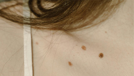
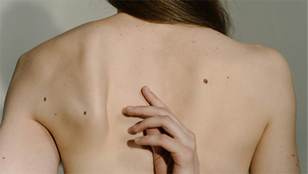
    How to Perform a Thorough At-Home Skin Self-Exam
    To effectively use the ABCDE rule, you must know your skin. We recommend performing a self-exam once a month in a well-lit room with a full-length mirror and a hand mirror.

    Examine the Front and Back: Use the full-length mirror to check your chest, stomach, and back. Don’t forget the areas under the breasts and in skin folds.

    Check the Extremities: Look at your forearms, palms, and the spaces between your fingers. Check your legs, the soles of your feet, and between your toes.

    Use a Hand Mirror for Hard-to-See Areas: Use a hand mirror to examine your scalp (parting your hair with a comb), the back of your neck, and your buttocks.

    Document Your Findings: If you find a spot that looks suspicious, take a clear, well-lit photo of it with a coin next to it for size reference. This helps your dermatologist track evolution over time.

    Why a Professional Exam is Different
    While the ABCDE rule is your first line of defense, it has limitations. Some types of skin cancer, like Amelanotic Melanoma, have no pigment at all and look like a harmless pink bump. Others might be hidden in places you can’t see, like behind the ears or on the scalp.

    At Chattanooga Skin and Cancer Clinic, we utilize Dermoscopy. This involves using a specialized handheld microscope that uses polarized light to see below the surface layer of the skin. This allows our residency-trained practitioners to see the “architecture” of a mole. By seeing how the pigment is structured, we can often determine if a mole is suspicious long before it would be obvious to the naked eye.

    The Tennessee Factor: Why Our Region is High-Risk
    Living in the Tennessee Valley brings unique risks. With our high UV index during the summer months and our community’s love for outdoor activities, from hiking the Cumberland Trail to boating on Chickamauga Lake, our skin is frequently exposed to damaging radiation.

    Cumulative sun exposure and occasional blistering sunburns both increase your risk of developing melanoma later in life. As a locally owned and independent clinic, we have seen firsthand how the Tennessee sun affects our neighbors. This is why we advocate so strongly for the “Yearly Skin Check” as a non-negotiable part of your healthcare routine.

    When to See a Dermatologist
    If you find a mole that meets even one of the ABCDE criteria, do not wait. Early-stage melanoma can often be treated with a simple in-office excision. However, if left to grow, the treatment becomes much more invasive.

    When you visit us, you aren’t just seeing a provider; you are seeing a team of board-certified dermatologists, nurse practitioners, and physician assistants who specialize exclusively in skin health. We provide expert care at every visit, ensuring that your concerns are heard and your skin is thoroughly evaluated.

    Conclusion: Your Skin, Our Mission
    Your skin is your body’s largest organ, and it works hard to protect you. The least you can do is keep an eye on it. By mastering the ABCDE rule and scheduling regular professional screenings, you are taking a massive step toward a long, healthy life.

    Chattanooga Skin and Cancer Clinic has been the trusted name in dermatology for over 50 years. Whether you have a suspicious mole or it’s simply time for your annual check-up, our family-owned practice is here to provide the expert, residency-trained care you deserve.

    Don’t wait for a “red flag” to become a problem. Contact us today at our Chattanooga, Cleveland, or Kimball offices to schedule your full-body skin examination.

  • Basal Cell vs Squamous Cell Carcinoma: What’s the Difference?

    Basal Cell vs Squamous Cell Carcinoma: What’s the Difference?

    Basal cell carcinoma and squamous cell carcinoma are the two most common types of skin cancer, and while both are highly treatable when caught early, they develop from different cell types, look different on the skin, and can behave in very different ways. Basal cell carcinoma (BCC) starts in the basal cells at the bottom of the outer skin layer, tends to grow slowly, and rarely spreads to other parts of the body. Squamous cell carcinoma (SCC) starts in the squamous cells closer to the skin’s surface, can grow faster, and carries a slightly higher risk of spreading if left untreated. According to the Skin Cancer Foundation, more than 4 million cases of BCC and over 1.8 million cases of SCC are diagnosed in the United States each year. Knowing how to tell these two cancers apart, and understanding when to get checked, can make a real difference in outcomes. At Chattanooga Skin and Cancer Clinic, our board-certified dermatologists diagnose and treat both types every day across our Chattanooga, Cleveland, and Kimball offices.

    How Do Basal Cell and Squamous Cell Carcinomas Develop Differently?

    Basal cell carcinoma forms in the deepest part of the epidermis, the outer layer of your skin. These cells are constantly dividing to produce new skin cells that push older ones toward the surface. When DNA damage (usually from UV exposure) causes basal cells to grow out of control, a BCC forms. Because these tumors grow slowly and tend to stay local, they are sometimes described as “less aggressive.” That said, left alone, a BCC can grow deep into surrounding tissue, damaging nerves, cartilage, and even bone.

    Squamous cell carcinoma originates higher up in the epidermis, in the flat cells that make up most of the skin’s outer surface. SCC often develops from precancerous spots called actinic keratoses, which are rough, scaly patches caused by years of sun exposure. While most SCCs stay localized, the risk of metastasis (spreading to lymph nodes or other organs) is higher than with BCC, particularly in tumors that are large, deep, or located on the ears, lips, or areas of chronic scarring.

    I find that patients are often surprised to learn these two cancers start just millimeters apart in the same layer of skin, yet their behavior can be so different.

    What Does Basal Cell Carcinoma Look Like on the Skin?

    BCC shows up in several forms, which can make it tricky to spot if you don’t know what to look for. The most common type appears as a pearly or waxy bump, often on the face, ears, or neck. It might have visible blood vessels running through it and a slightly translucent quality, almost like a small blister that never quite goes away.

    Other BCCs look like flat, flesh-colored or brown lesions on the chest or back. Some develop a central sore that crusts over, heals partially, then opens up again. This cycle of scabbing and reopening is one of the most reliable warning signs. A sore that refuses to fully heal within three to four weeks deserves a professional evaluation.

    Less common variants include morpheaform BCC, which looks like a pale, waxy scar and can be harder to detect because it blends into surrounding skin. These tend to have less defined borders, which makes them more challenging to treat.

    What Does Squamous Cell Carcinoma Look Like?

    SCC tends to look rougher and more textured than BCC. A common presentation is a firm, red nodule, often on sun-exposed areas like the face, ears, neck, hands, or forearms. The surface may be scaly or crusted, and the lesion can feel tender to the touch.

    Some SCCs appear as flat, reddish patches with an irregular border that slowly grow over time. Others develop into open sores that bleed or crust and don’t resolve on their own. On the lips, SCC can look like a persistent rough or cracked patch that doesn’t respond to lip balm or chapstick.

    One thing to keep in mind: SCC can also develop in areas that don’t get much direct sun, including the genitals, inside the mouth, and on scars or chronic wounds. People with weakened immune systems, including organ transplant recipients, are at higher risk for these less typical presentations.

    What Are the Main Risk Factors for Each Type?

    UV exposure is the single biggest risk factor for both BCC and SCC. That includes natural sunlight and tanning beds. People with fair skin, light eyes, and a history of sunburns are at the highest risk, though skin cancer can develop in people of every skin tone.

    For BCC specifically, intermittent intense sun exposure (think weekend sunburns or vacation burns) plays a bigger role. Childhood sunburns are especially relevant. BCC also tends to show up on areas with the most cumulative sun exposure, primarily the face, head, and neck.

    SCC, on the other hand, is more closely tied to cumulative, long-term sun exposure. People who work outdoors or spend considerable time in the sun over decades have an elevated risk. Other SCC risk factors include a history of precancerous actinic keratoses, previous radiation therapy, chronic wounds, and immunosuppression.

    Age is a factor for both types. Diagnoses become much more common after age 50, though dermatologists are increasingly seeing both BCC and SCC in younger patients, likely tied to tanning bed use and increased recreational UV exposure.

    How Are Basal Cell and Squamous Cell Carcinomas Treated?

    Treatment depends on the type, size, location, and depth of the cancer. For small, superficial BCCs, options include surgical excision (cutting out the tumor with a margin of healthy tissue), curettage and electrodesiccation (scraping the tumor and using an electric needle to destroy remaining cells), or topical medications like imiquimod or fluorouracil that stimulate the immune system to attack abnormal cells.

    For larger or more complex BCCs, and for most SCCs, Mohs micrographic surgery is considered the gold standard. During Mohs surgery, the surgeon removes thin layers of tissue one at a time, examining each layer under a microscope immediately. This process continues until no cancer cells remain. Mohs offers the highest cure rate (up to 99% for primary BCCs and up to 97% for primary SCCs, according to the American College of Mohs Surgery) while preserving as much healthy tissue as possible.

    At Chattanooga Skin and Cancer Clinic, we perform Mohs surgery at both our Chattanooga and Cleveland locations. For patients at our Kimball office, we coordinate referrals to one of these locations when Mohs is the best option.

    SCC that has spread past the skin may require additional treatments such as radiation therapy, systemic chemotherapy, or immunotherapy. These cases are less common, but they reinforce why early detection matters so much.

    Why Does Early Detection Make Such a Big Difference?

    When caught early, both BCC and SCC have excellent prognosis. A BCC removed in its early stages almost never comes back and almost never spreads. The same is true for most early-stage SCCs. The trouble starts when people wait.

    A BCC that grows unchecked for years can invade bone and cartilage, requiring complex reconstructive surgery. An SCC that isn’t addressed can metastasize, and metastatic SCC has a five-year survival rate of about 34%, according to the American Cancer Society. That’s a sharp contrast from the nearly 100% cure rate when it’s caught and treated locally.

    The takeaway is simple: if something on your skin is new, changing, or not healing, get it looked at. A 15-minute skin check could save you from a far more complicated treatment down the road.

    How Can You Reduce Your Risk of Both Types of Skin Cancer?

    Sun protection is the most effective prevention strategy for both BCC and SCC. That means broad-spectrum sunscreen with SPF 30 or higher applied every two hours when outdoors, protective clothing (hats, long sleeves, UV-blocking sunglasses), and seeking shade during peak UV hours between 10 a.m. and 4 p.m. Tanning beds should be avoided entirely. The World Health Organization classifies UV-emitting tanning devices as Group 1 carcinogens, the same category as tobacco.

    Regular skin self-exams help you spot changes early. Once a month, check your entire body in a well-lit room with a full-length mirror. Use a hand mirror for hard-to-see areas like your back, scalp, and the soles of your feet. If you notice anything new or different, a firm bump, a scaly patch, a sore that won’t heal, schedule an appointment.

    Annual professional skin exams are also a smart habit, especially if you have a personal or family history of skin cancer, fair skin, a history of extensive sun exposure, or a large number of moles. Our dermatologists at Chattanooga Skin and Cancer Clinic perform thorough head-to-toe screenings designed to catch problems before they become serious.

    Frequently Asked Questions About Basal Cell and Squamous Cell Carcinoma

    Can basal cell or squamous cell carcinoma be fatal?

    BCC is almost never fatal. It grows slowly and very rarely spreads past the original site. SCC carries a low but real risk of metastasis, particularly when left untreated for a long time or when it occurs in immunocompromised patients. Early treatment for both types is nearly always curative.

    What is Mohs surgery and when is it recommended?

    Mohs micrographic surgery removes skin cancer one layer at a time, with each layer examined under a microscope before the next is removed. It’s recommended for cancers in cosmetically or functionally sensitive areas (face, ears, hands), for large or recurrent tumors, and for aggressive subtypes. It offers the highest cure rates with the smallest possible wound.

    Where can I get a skin cancer screening near Chattanooga?

    Chattanooga Skin and Cancer Clinic provides full-body skin cancer screenings at all three locations: Chattanooga (6061 Shallowford Road), Cleveland (3891 Adkisson Drive), and Kimball (400 Dixie Lee Center Rd). Appointments are available Monday through Friday. Call 423-899-2700 for the Chattanooga office, 423-479-8648 for Cleveland, or 423-815-9975 for Kimball.

  • What to Expect at Your First Full-Body Skin Exam

    What to Expect at Your First Full-Body Skin Exam

    A full-body skin exam is a head-to-toe visual screening performed by a dermatologist to check for skin cancer, precancerous spots, and other skin concerns. The exam itself takes about 10 to 15 minutes, is painless, and requires no special preparation. Your dermatologist will examine your entire skin surface, including areas you can’t easily see yourself, like your scalp, back, and the soles of your feet. According to the American Academy of Dermatology, skin cancer is the most common cancer in the United States, with an estimated one in five Americans developing it in their lifetime. Early detection through routine screening is one of the most effective ways to improve outcomes. At Chattanooga Skin and Cancer Clinic, our board-certified, residency-trained dermatologists have been performing these exams for over 50 years, and we see roughly 250 patients per day across our three locations in Chattanooga, Cleveland, and Kimball.

    Why Should You Get a Full-Body Skin Exam?

    Skin cancer is common, but it’s also one of the most treatable cancers when found early. The problem is that many skin cancers don’t hurt, itch, or cause obvious symptoms in their earliest stages. A small basal cell carcinoma might look like a pimple that never quite clears up. An early melanoma might resemble an ordinary mole. Without a trained eye examining your skin systematically, these early signs can go unnoticed for months or even years.

    Professional skin exams catch things self-checks miss. A 2016 study published in the Journal of the American Academy of Dermatology found that dermatologists identified melanomas that were thinner (and therefore more treatable) compared to melanomas first noticed by patients themselves. Thinner melanomas have dramatically better survival rates, so catching them at that stage makes a measurable difference.

    I think most people are surprised by how quick the exam is relative to the peace of mind it provides. Ten minutes once a year is a small investment for something that could catch a serious problem before it becomes complicated.

    How Should You Prepare for a Skin Exam?

    There isn’t much you need to do. Show up with clean skin and remove any nail polish from your fingers and toes a day or two beforehand, since melanoma can develop under the nails. Skip heavy makeup on the day of your appointment so your dermatologist can see your facial skin clearly.

    Take a few minutes before your visit to note anything that concerns you: a mole that changed color or shape, a spot that bleeds or crusts, a patch of rough skin that won’t go away. Write these down or take a photo. Having specific concerns ready helps your dermatologist prioritize areas during the exam and gives them context they wouldn’t otherwise have.

    You don’t need a referral to book a skin exam at most dermatology practices, including ours. If you have a family history of skin cancer or melanoma, mention that when you schedule so it’s noted in your chart before you arrive.

    What Happens During the Exam Itself?

    When you arrive, a medical assistant will bring you to an exam room and ask you to change into a gown. If you’re uncomfortable with a full exam, you can ask to keep undergarments on, but it’s worth knowing that skin cancer can develop anywhere on the body, including areas covered by clothing. Your dermatologist will work with whatever level of coverage you’re comfortable with.

    The exam follows a systematic, head-to-toe approach. Your dermatologist will start at the scalp, parting your hair to examine the skin beneath. They’ll move to the face, ears (front and back), neck, and down through the chest, abdomen, arms, hands (including between the fingers and under the nails), back, legs, feet, and the soles of your feet. They’ll use a dermatoscope, a handheld magnifying device with a built-in light, to get a closer look at moles or spots that need more detailed evaluation.

    The whole process usually wraps up in 10 to 15 minutes. It doesn’t involve any needles, blood draws, or discomfort. Your dermatologist may ask you questions during the exam: “How long have you had this mole?” or “Has this spot changed recently?” These aren’t trick questions. They’re gathering context to make better clinical decisions.

    What Happens If the Dermatologist Finds Something?

    Finding “something” doesn’t automatically mean cancer. Dermatologists evaluate dozens of spots during a typical exam, and the vast majority turn out to be benign moles, seborrheic keratoses (harmless age spots), or minor irritations. If your dermatologist sees a lesion that looks suspicious, they may recommend a biopsy.

    A skin biopsy is a quick, in-office procedure. After numbing the area with a local anesthetic, the dermatologist removes a small sample of tissue (sometimes the entire spot, sometimes just a portion) and sends it to a pathology lab for analysis. You’ll typically get results within one to two weeks. If the biopsy confirms cancer or a precancerous condition, your dermatologist will walk you through treatment options in detail, including what the procedure involves, expected recovery time, and what follow-up looks like.

    At Chattanooga Skin and Cancer Clinic, if a biopsy reveals a cancer that would benefit from Mohs micrographic surgery, we can perform that procedure in-house at our Chattanooga and Cleveland offices. Keeping diagnosis and treatment under the same roof reduces the number of appointments and the gap between finding a problem and fixing it.

    Does a Full-Body Skin Exam Hurt?

    No. The exam itself is completely painless. Your dermatologist is looking at your skin, not poking, prodding, or pressing on it. The dermatoscope rests lightly against the skin for a few seconds at a time when evaluating specific spots, but there’s no discomfort involved.

    If a biopsy is needed, the numbing injection feels like a brief pinch, similar to a small bee sting that lasts about two seconds. After that, you won’t feel the biopsy itself. Most patients say the anticipation was worse than the actual experience.

    How Often Should You Get a Full-Body Skin Exam?

    For most adults, an annual skin exam is a solid baseline. If you have a higher risk profile, your dermatologist may recommend more frequent screenings, such as every six months. Higher-risk categories include people with a personal history of skin cancer, a family history of melanoma, a large number of moles (more than 50), a history of blistering sunburns, fair skin with light eyes and hair, or a weakened immune system.

    Your dermatologist will tell you at the end of your exam how often they’d like to see you. Follow their recommendation. Skin cancer caught at a routine follow-up visit is almost always easier and less expensive to treat than one that’s been growing undetected.

    What Should You Do Between Exams?

    Monthly self-exams are the best habit you can build between annual screenings. Stand in front of a full-length mirror in a well-lit room and check yourself from head to toe. Use a hand mirror for your back, scalp, and other hard-to-see areas. You’re looking for the “ABCDEs” of melanoma: Asymmetry (one half doesn’t match the other), Border irregularity (edges are ragged or blurred), Color variation (multiple shades of brown, black, red, or white in a single spot), Diameter (larger than a pencil eraser, about 6mm), and Evolution (any change in size, shape, or color over time).

    Outside of melanoma, keep an eye out for any new growths, sores that won’t heal, or patches of rough or scaly skin that persist. Not every new spot is skin cancer, not even close, but changes that don’t resolve on their own within a few weeks are worth a professional opinion.

    Sun protection between exams also matters. Broad-spectrum sunscreen (SPF 30+), protective clothing, hats, and staying out of direct midday sun all reduce your risk of developing new skin cancers. Prevention and early detection work best as a team.

    What If You Feel Nervous or Embarrassed About the Exam?

    This is completely normal, and dermatologists know it. A full-body skin exam involves being seen in a gown, and that’s a vulnerable position for anyone. Here’s the reality: your dermatologist does this dozens of times a day. They’re focused on your skin, not on anything else. The exam room is private, and you can ask to have a nurse or medical assistant present if that makes you more comfortable.

    If there are specific areas of your body you’re not comfortable having examined, say so. Your dermatologist would rather examine most of your skin than have you skip the appointment entirely. You can also wear underwear during the exam and simply mention the areas you’d like them to skip. Some screening is always better than none.

    First-time patients at our clinic often tell us afterward that the exam was much less awkward than they expected. The anticipation is almost always the hardest part.

    Frequently Asked Questions About Full-Body Skin Exams

    Do I need a referral to see a dermatologist for a skin exam?

    In most cases, no. Many insurance plans allow you to book directly with a dermatologist without a referral from your primary care physician. Check with your insurance provider or call our office to verify your specific plan. Chattanooga Skin and Cancer Clinic accepts most major insurance plans.

    Can skin cancer develop in places that don’t get sun exposure?

    Yes. While UV exposure is the primary risk factor, skin cancer can develop on the soles of the feet, under the nails, on the scalp, and in the genital area. This is one of the reasons a full-body exam covers all areas, not just sun-exposed skin.

    Where can I schedule a full-body skin exam near Chattanooga?

    Chattanooga Skin and Cancer Clinic offers full-body skin exams at all three locations. Our Chattanooga office is at 6061 Shallowford Road (423-899-2700), our Cleveland office is at 3891 Adkisson Drive (423-479-8648), and our Kimball office is at 400 Dixie Lee Center Rd (423-815-9975). Appointments are available Monday through Friday.