Category: Kimball

  • The 4 Types of Rosacea Explained: Which Type Do You Have?

    The 4 Types of Rosacea Explained: Which Type Do You Have?

    The 4 Types of Rosacea Explained: Which Type Do You Have?

    Close-up of a patient's cheek with hyperpigmentation and sun damage, showcasing skin rejuvenation concerns
    Spotting the early signs of sun damage or uneven texture? Our Kimball skincare experts can help restore your skin’s natural glow.

    Rosacea is one of the most misunderstood skin conditions out there. A lot of people spend years treating what they think is adult acne, dry skin, or sun damage before a dermatologist finally connects the dots. The redness keeps coming back. The skin feels sensitive no matter what products you use. Something is clearly going on, but the label stays elusive. If that sounds familiar, there is a good chance rosacea is the reason.

    What makes rosacea especially tricky is that it does not look the same on every person. Some people develop flushing and broken blood vessels. Others deal with acne-like breakouts. Some have thickened skin, and others struggle with eye irritation that they would never connect to a skin condition. Rosacea has four distinct subtypes, and understanding which one you have is the starting point for getting it properly managed.

    According to the National Rosacea Society, more than 16 million Americans are affected by rosacea, and many of them do not realize they have it. This article breaks down each of the four types, what they look like, how they differ, and what you can do about them.


    What Is Rosacea and Why Does It Come in Different Types?

    Rosacea is a chronic skin condition that causes redness, inflammation, and a range of other symptoms primarily on the face. It tends to cycle through flares and calmer periods, often triggered by heat, sun, spicy food, alcohol, stress, or certain skincare products. There is no cure, but with the right treatment plan, most people can keep it well under control.

    The reason rosacea has subtypes is that it affects different structures of the skin in different ways. The blood vessels near the surface, the oil glands, the connective tissue, and even the eyes can all be involved depending on the person. Doctors and researchers use a subtype classification system to describe these patterns and match them to the most effective treatments.

    It is worth knowing that subtypes are not always mutually exclusive. Some people experience two or more types at once, or see one subtype evolve into another over time if the condition goes untreated. That is another reason early diagnosis from a board-certified dermatologist matters so much.


    What Is Subtype 1 and How Does Erythematotelangiectatic Rosacea Show Up?

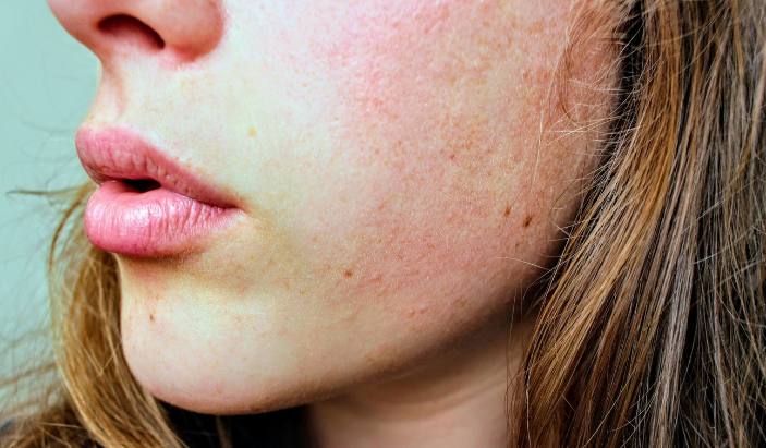
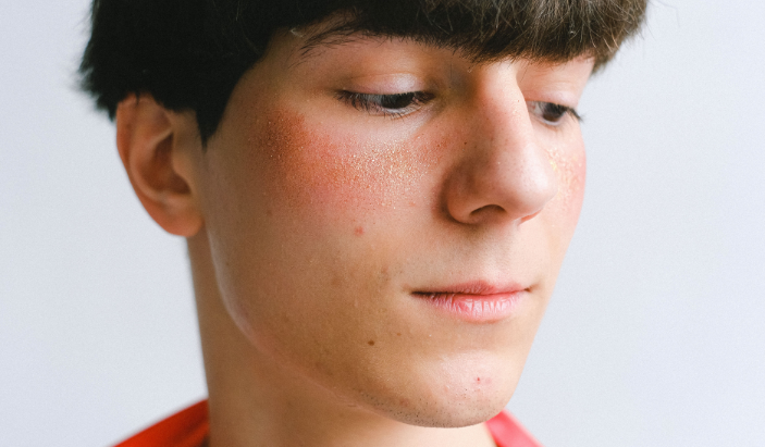
    Subtype 1 is called erythematotelangiectatic rosacea, often abbreviated as ETR. The name is a mouthful, but the presentation is fairly straightforward: persistent redness across the cheeks, nose, chin, or forehead, often accompanied by flushing and visible blood vessels close to the skin’s surface.

    People with ETR frequently report that their face feels like it is burning or stinging, even without an obvious trigger. The skin tends to be sensitive and reactive, flaring up in response to temperature changes, exercise, wind, hot beverages, or sun exposure. The redness does not fade the way a normal flush would. It lingers, and over time, the tiny blood vessels known as telangiectasias become more visible, creating a web of fine red or pink lines across the face.

    This subtype is the most commonly recognized form of rosacea. Many people with ETR try to treat the redness with moisturizers or over-the-counter products, but those rarely address what is actually happening beneath the surface. A dermatologist can prescribe topical medications that reduce redness or recommend in-office laser and light treatments that target the visible blood vessels directly.


    What Is Subtype 2 and How Is Papulopustular Rosacea Different From Acne?

    Young male patient experiencing facial redness and rosacea-like flushing on the cheeks, illustrating sensitive skin treatments.
    Persistent facial flushing or redness can be managed with a custom treatment plan at our Kimball clinic.

    Subtype 2 is papulopustular rosacea, and it is the one most frequently mistaken for acne. It causes red bumps called papules and pus-filled blemishes called pustules, usually concentrated in the central face. That combination of breakouts plus background redness is the classic picture of this subtype.

    The key difference between papulopustular rosacea and acne comes down to what you do not see. Rosacea does not produce blackheads or whiteheads. Acne does. If you have what looks like adult acne but there are no blackheads in the mix, rosacea becomes a much more likely explanation. Age is another clue. Papulopustular rosacea is more common in adults, particularly women in their 30s, 40s, and 50s.

    Using standard acne treatments on this subtype can backfire badly. Many acne products contain ingredients like benzoyl peroxide or retinoids that are simply too harsh for rosacea-prone skin. They strip and irritate the skin barrier, making the redness and breakouts worse. A proper diagnosis is the only way to avoid that cycle. A dermatologist can confirm which condition you are dealing with and prescribe treatments like topical azelaic acid, metronidazole, or oral antibiotics that are designed for rosacea specifically.


    What Is Subtype 3 and Who Does Phymatous Rosacea Typically Affect?

    Subtype 3 is phymatous rosacea, the rarest and most dramatic presentation of the condition. It causes a thickening and irregular texture of the skin due to the enlargement of oil glands and a buildup of connective tissue. The nose is the most commonly affected area, and when it progresses significantly, the condition is called rhinophyma.

    Rhinophyma creates a bulbous, bumpy appearance on the nose, with enlarged pores and a rough texture. It can also affect the chin, forehead, ears, and eyelids, though this is less frequent. Phymatous rosacea is much more common in men than in women, and the reasons for that are not entirely clear, though hormonal factors are thought to play a role.

    This subtype tends to develop gradually over years, often in people who had untreated rosacea for a long time. That makes it one of the stronger arguments for getting rosacea evaluated and managed early. Once phymatous changes develop, they do not reverse on their own. Treatment in the earlier stages involves prescription medications to slow progression. More advanced cases may require laser resurfacing or surgical intervention to reshape the tissue.


    What Is Subtype 4 and How Does Ocular Rosacea Affect the Eyes?

    Subtype 4 is ocular rosacea, and it is the most overlooked type because many people do not connect eye symptoms to a skin condition. Ocular rosacea affects the eyes and the skin around them, causing redness, irritation, dryness, and a persistent gritty or burning sensation. The eyelids may become swollen, crusty, or inflamed, a condition called blepharitis.

    Some people with ocular rosacea develop it alongside one of the other subtypes. Others experience the eye symptoms before any skin changes appear, which makes diagnosis particularly confusing. Left unmanaged, ocular rosacea can lead to sensitivity to light, blurred vision, and in more serious cases, corneal damage.

    Treatment for ocular rosacea often involves a combination of approaches: warm compresses, lid hygiene routines, prescription eye drops, and in some cases oral antibiotics that reduce inflammation. A dermatologist and an ophthalmologist sometimes work together to manage this subtype, especially when both skin and eye symptoms are present.


    How Do You Know Which Type of Rosacea You Have?

    Identifying your subtype is not something you can do reliably on your own, and trying to piece it together from an internet search can lead you in the wrong direction. A board-certified dermatologist evaluates your skin in person, considers your symptom history, and identifies not just which subtype is present but whether multiple subtypes are overlapping.

    The distinction matters because treatment varies meaningfully by subtype. The approach for ETR focuses on reducing vascular reactivity and protecting a sensitive skin barrier. Papulopustular rosacea calls for anti-inflammatory treatments. Phymatous changes may require procedures. Ocular involvement often needs specialized eye care in addition to dermatology management. A one-size approach simply does not work across all four.

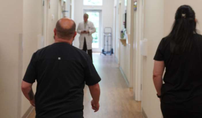
    At Chattanooga Skin and Cancer Clinic, board-certified dermatologists have been evaluating and treating rosacea patients across the Chattanooga, Cleveland, and Kimball area since 1973. With three convenient locations and same-team continuity of care, patients get a thorough assessment and a treatment plan that fits their specific presentation.


    What Triggers Make Rosacea Worse Across All Four Types?

    Regardless of subtype, rosacea responds to a similar set of triggers. Sun exposure is one of the most common, and Tennessee summers can be particularly rough on rosacea-prone skin. UV radiation dilates blood vessels and causes lasting inflammation over time. Daily broad-spectrum sunscreen is a non-negotiable part of managing any type of rosacea.

    Heat, spicy foods, alcohol (particularly red wine), hot beverages, stress, and intense exercise all commonly provoke flares. So do certain skincare ingredients: fragrances, alcohol-based toners, witch hazel, and harsh exfoliants. Keeping a simple trigger diary helps identify personal patterns, since not everyone reacts to the same things.

    The goal of trigger management is not to eliminate every possible irritant from your life. It is to identify your top offenders so you can minimize them where it makes a real difference. Combined with prescription treatment from a dermatologist, trigger awareness gives patients the best shot at keeping rosacea calm long-term.


    Frequently Asked Questions About the Types of Rosacea

    Medical staff and dermatologist walking down the hallway of Chattanooga Skin and Cancer Clinic, a premier dermatology clinic.
    Our expert dermatology team in Kimball working behind the scenes to bring you personalized, medical-grade skin solutions

    Can you have more than one type of rosacea at the same time?

    Yes. It is fairly common for people to experience two subtypes simultaneously. Subtype 1 and Subtype 2 often occur together, with persistent redness and visible blood vessels accompanying acne-like breakouts. Ocular symptoms can also appear alongside any of the skin-based subtypes.

    Does rosacea subtype change over time?

    It can. Rosacea is a progressive condition in many people, meaning it tends to worsen if left untreated. Subtype 1 can evolve to include the papules and pustules of Subtype 2 over time. Phymatous changes typically develop after years of chronic, unmanaged inflammation. Getting diagnosed and starting treatment early is the best way to slow or prevent that progression.

    Is rosacea more common in certain skin tones?

    Rosacea is most frequently diagnosed in people with fair skin, but it affects people of all skin tones. On medium to deeper skin tones, it often goes unrecognized because the classic redness is harder to see. Symptoms like burning, stinging, bumps, or eye irritation may still be present, and a dermatologist familiar with rosacea presentations across diverse skin tones can make an accurate diagnosis.

    Are there rosacea treatments that work for all four types?

    Some treatments overlap across subtypes, particularly topical and oral anti-inflammatory medications. That said, the most effective treatment plans are specific to the subtype. Laser treatments that target blood vessels are most useful for Subtype 1. Antibiotic therapies are more central to Subtype 2. Subtype 3 may require procedural intervention. Subtype 4 often needs targeted eye care. Treatment works best when it is matched to what is actually happening in your skin.

    When should you see a dermatologist about rosacea?

    As soon as you suspect it. Rosacea does not improve on its own, and the longer it goes unmanaged, the more ingrained the changes to the skin can become. If you have persistent facial redness, recurring breakouts without blackheads, skin that feels easily irritated, or any eye discomfort that your eye doctor has not fully explained, a dermatology evaluation is the right move. Early treatment produces the best long-term results.


    Ready to Get a Clear Answer About Your Skin?

    Living with rosacea is manageable, but only once you understand what you are actually dealing with. Guessing at a diagnosis and cycling through the wrong products wastes time and often makes the condition worse. The four subtypes of rosacea are distinct enough that proper identification genuinely changes the treatment path.

    The team at Chattanooga Skin and Cancer Clinic includes board-certified dermatologists who have spent decades treating rosacea patients across Southeast Tennessee. Appointments are available in Chattanooga, Cleveland, and Kimball. Call the location nearest you or request an appointment online at chattskinandcancer.com.

  • Eczema in Babies and Toddlers: What Parents Need to Know

    Eczema in Babies and Toddlers: What Parents Need to Know


    Your baby wakes up again at 2 a.m., scratching at the same raw patches of skin on their cheeks and elbows. You’ve tried switching detergents, avoiding certain foods, and slathering on whatever lotion was at the top of the search results. Nothing is really working, and the pediatrician used a word you’ve been reading about ever since: eczema. If that scenario sounds familiar, you are not alone. Eczema is one of the most common skin conditions in young children, and it is also one of the most misunderstood.

    Eczema in babies and toddlers affects roughly 10 to 20 percent of children in the United States, according to the National Eczema Association. It tends to show up in the first year of life, often before a baby’s first birthday. For many parents, the diagnosis raises more questions than answers. What exactly is eczema? What causes it to flare? How do you manage it day to day, and when does a child need to see a dermatologist? This article answers all of those questions clearly and practically, so parents in the Chattanooga area and beyond can feel confident taking care of their child’s skin.


    What Is Eczema and Why Does It Affect So Many Babies?

    A crying infant showing signs of facial discomfort and skin sensitivity from baby eczema.
    Severe itching and skin discomfort from infant eczema can deeply disrupt a baby’s comfort and sleep.

    Eczema, medically known as atopic dermatitis, is a chronic inflammatory skin condition where the skin’s protective barrier does not function properly. A healthy skin barrier locks in moisture and keeps irritants and allergens out. In children with eczema, that barrier is compromised, meaning the skin loses water more easily and becomes vulnerable to environmental triggers.

    The result is skin that is dry, itchy, and prone to red, inflamed patches. In babies, these patches often appear on the face, especially the cheeks and forehead. As children grow into toddlers, eczema tends to shift to the elbow creases, behind the knees, and around the wrists. The itching can be intense, and because babies and toddlers cannot stop themselves from scratching, the skin can break open, which raises the risk of infection.

    Eczema is not contagious. It is not caused by poor hygiene or anything a parent did wrong. Genetics play a large role. Children with a parent or sibling who has eczema, asthma, or seasonal allergies are significantly more likely to develop it themselves. This connection, called the atopic march, means that early eczema in infants can sometimes be a predictor of later allergies or asthma.


    How Do You Know If Your Baby Has Eczema?

    Eczema can look different depending on the child’s age and skin tone. In lighter skin, inflamed patches appear red or pink. In deeper skin tones, eczema may look more purple, brown, or grayish, which leads to it being missed or misdiagnosed more often in children of color.

    Common signs to watch for in babies under 12 months include dry, scaly patches on the cheeks, scalp, or forehead; oozing or crusting skin; and visible discomfort, especially when the baby is warm or after a bath. Toddlers often show eczema in the skin folds at the elbows, wrists, knees, and ankles. The skin may appear thickened or leathery in areas that have been scratched repeatedly over time.

    One hallmark of eczema is the cycle of flares and relative calm. A child might have clear skin for weeks, then break out after a trigger like cold weather, sweating, or exposure to a new product. That unpredictable pattern can make management feel frustrating, but understanding triggers is one of the most useful tools parents have.


    What Triggers Eczema Flares in Young Children?

    Triggers vary from child to child, but some of the most consistent culprits include dry air, sweat, synthetic fabrics, certain soaps and fragrances, dust mites, pet dander, and stress. In the Southeast, where Chattanooga’s climate includes both humid summers and cold winters, children with eczema can face year-round challenges as the environment shifts.

    Food allergies sometimes overlap with eczema, which leads many parents to assume that diet is the root cause. The relationship between eczema and food is real but complicated. The American Academy of Dermatology notes that food allergies are found in about 30 percent of children with moderate to severe eczema, with milk, eggs, wheat, soy, and peanuts being the most common culprits. Eliminating foods without guidance can be harmful to a young child’s nutrition. A board-certified dermatologist or allergist should evaluate whether food is genuinely contributing to a child’s flares before any dietary changes are made.

    Temperature is another major factor. Overheating causes sweating, which irritates the skin. Keeping bedrooms cool, dressing children in loose cotton layers, and avoiding heavy blankets at night can all reduce the frequency of flares.


    How Is Eczema in Babies and Toddlers Treated?

    There is no cure for eczema, but it is very manageable with a consistent routine. The foundation of eczema care for young children is moisturizing frequently and effectively. Applying a thick, fragrance-free cream or ointment immediately after a bath, while the skin is still slightly damp, helps lock in moisture before it evaporates. Creams and ointments outperform thin lotions for this purpose because they contain more lipid content and create a stronger barrier.

    Bathing in lukewarm water for five to ten minutes, then patting the skin dry before applying moisturizer, is a routine that dermatologists recommend consistently. Hot water strips the skin’s natural oils and worsens dryness. Fragrance-free, gentle cleansers should replace traditional soaps.

    When moisturizing alone does not control flares, a pediatric dermatologist may recommend topical corticosteroids. These prescription creams reduce inflammation and are safe when used correctly. Parents are often cautious about steroids, and that concern is understandable, but low-potency topical steroids used as directed on affected areas are a well-established treatment option. A dermatologist will choose the appropriate strength for the child’s age and the area being treated.

    Newer non-steroidal prescription treatments, such as topical calcineurin inhibitors, are also available for children who need ongoing management without repeated steroid use. For more severe cases, a pediatric dermatologist may discuss systemic treatments or biologic medications. Dupilumab received FDA approval for children as young as six months old in 2023, marking a meaningful step forward in treating moderate to severe eczema in infants.


    When Should You See a Dermatologist Instead of Waiting It Out?

    Pediatricians are a good starting point for mild eczema, but there are clear situations where a dermatologist’s expertise makes a real difference. A board-certified dermatologist has specialized training in skin conditions that goes beyond what most general practitioners receive.

    See a dermatologist if your child’s eczema is not improving after two to four weeks of consistent moisturizing and over-the-counter care. Seek care sooner if the skin appears infected, which may look like crusting, honey-colored discharge, increased redness, warmth, or swelling. Scratched eczema skin is an open door for bacterial infections, particularly staph, and those infections need prompt treatment.

    A dermatologist can also confirm the diagnosis when eczema looks similar to other conditions like psoriasis, seborrheic dermatitis, or contact dermatitis. Getting the right diagnosis matters because these conditions respond to different treatments. Treating one condition like another will not produce the results the child needs.

    Chattanooga Skin and Cancer Clinic has been caring for patients of all ages since 1973, including infants and toddlers with chronic skin conditions. All providers are board-certified dermatologists with experience in pediatric dermatology. With three locations across Chattanooga, Cleveland, and Kimball, families across Southeast Tennessee can access specialized care close to home.


    What Daily Habits Make the Biggest Difference for Kids with Eczema?

    A parent checking a baby's back for dry patches and toddler eczema flares in Chattanooga.
    Consistently checking your child’s skin folds and back helps track eczema triggers and flare-ups early.

    Consistency is everything with eczema management. Moisturizing once a day is helpful; moisturizing twice a day is better. The goal is to keep the skin barrier as intact as possible so it has less opportunity to react to triggers.

    Clothing choices matter more than most parents realize. Synthetic fabrics like polyester and nylon trap heat and create friction against the skin. Loose-fitting, 100 percent cotton clothing lets the skin breathe and reduces irritation. Wash new clothing before the first wear to remove manufacturing residues, and use fragrance-free, dye-free detergents consistently.

    Keeping a simple log of flares can help parents and dermatologists identify patterns. Note what the child ate, what products were used, changes in weather or environment, and how the skin responded. Over time, these records often reveal connections that are not obvious in the moment.

    Short, trimmed fingernails reduce the skin damage that comes from scratching during sleep. For infants who scratch at night, lightweight cotton mittens can protect the skin until they are old enough to understand not to scratch.


    Does Eczema in Babies Go Away on Its Own?

    Many children do experience significant improvement as they get older. Studies suggest that about 50 percent of children with infant eczema see it resolve or become much milder by age six. Another portion will continue to have flares into adolescence or adulthood, though often less severely than in early childhood.

    The trajectory is hard to predict for any individual child. Children with more severe early eczema, or those who also have food allergies or asthma, are more likely to have persistent symptoms. Early, consistent treatment and close management with a dermatologist give children the best chance at keeping flares minimal and protecting skin health long-term.

    Parents should also know that eczema does not reflect on their parenting. Many children with eczema go on to have completely normal, active childhoods when their condition is well managed.


    Frequently Asked Questions About Eczema in Babies and Toddlers

    Is eczema the same as dry skin?

    No. While dry skin is a feature of eczema, eczema is a chronic inflammatory condition with a genetic component. Simple dry skin responds to moisturizer and does not cause the chronic, itchy, inflamed patches that eczema does. If moisturizing alone is not controlling the problem, the skin condition likely needs a dermatologist’s assessment.

    Can breastfeeding or formula affect my baby’s eczema?

    There is some evidence that breastfeeding may offer a modest protective effect against developing eczema, but breastfeeding does not prevent it entirely and formula does not cause it. If a nursing mother suspects her diet is affecting her baby’s skin, speaking with a dermatologist or allergist is the right path forward rather than eliminating foods without guidance.

    Are topical steroids safe to use on a baby’s skin?

    Low-potency topical corticosteroids are considered safe for short-term use in infants and toddlers when prescribed by a dermatologist and applied as directed. Concerns about steroid side effects are most relevant when high-potency creams are overused for long periods. A board-certified dermatologist prescribes the appropriate strength and gives clear instructions on duration and frequency.

    What is the best moisturizer for baby eczema?

    Thick, fragrance-free creams or ointments tend to work better than thin lotions. Products like plain petroleum jelly, CeraVe Healing Ointment, or Vanicream Moisturizing Ointment are frequently recommended because they are fragrance-free and effective. The best moisturizer is one that is applied consistently and immediately after bathing while the skin is still slightly damp.

    When should I bring my toddler to Chattanooga Skin and Cancer Clinic for eczema?

    A toddler holding a stuffed animal while being examined at Chattanooga Skin and Cancer Clinic.
    Board-certified pediatric dermatologists offer advanced treatment plans for moderate to severe childhood eczema.

    Bring your child in if the eczema is not responding to at-home care after two to four weeks, if you see signs of infection, if the condition is affecting your child’s sleep or quality of life, or if you are unsure whether the diagnosis is correct. The practice’s board-certified dermatologists see patients across all age groups, including infants, at locations in Chattanooga, Cleveland, and Kimball. You can call the Chattanooga office at 423-899-2700 or the Cleveland office at 423-479-8648 to schedule an appointment.


    Getting Your Child the Right Care

    Eczema in babies and toddlers can feel relentless, especially in those early months when everything is new and sleep is already scarce. The good news is that with the right routine, the right triggers identified, and the right medical support in place, most children with eczema live comfortably and thrive.

    Chattanooga Skin and Cancer Clinic has been part of this community for more than 50 years, founded in 1973 and now serving patients at three locations across Southeast Tennessee. Every provider is board-certified, and the clinic treats patients from infancy through adulthood. If your baby or toddler is struggling with eczema and you are ready to move past guesswork, the team at Chattanooga Skin and Cancer Clinic is ready to help.

    Call 423-899-2700 for the Chattanooga location at 6061 Shallowford Road, 423-479-8648 for Cleveland at 3891 Adkisson Drive, or 423-815-9975 for the Kimball office at 400 Dixie Lee Center Rd. All three offices are open Monday through Friday, and new patient appointments are available.


  • How to Do a Self-Skin Check at Home (and What to Look For)

    How to Do a Self-Skin Check at Home (and What to Look For)

    A monthly self-skin check is one of the simplest things you can do to catch skin cancer early, and it takes about 10 minutes. You stand in front of a mirror, examine your skin from head to toe, and look for anything new, changing, or unusual. That’s it. No special equipment required. The reason this matters is straightforward: skin cancer is the most common cancer in the United States, affecting one in five Americans in their lifetime according to the American Academy of Dermatology. When caught early, most skin cancers are highly treatable. When caught late, treatment becomes more complex and outcomes get worse. The gap between early and late detection often comes down to whether someone was paying attention. A self-skin check fills the months between your annual professional skin exams and gives you a baseline understanding of your own skin, so when something does change, you notice. At Chattanooga Skin and Cancer Clinic, our board-certified dermatologists encourage every patient to build this habit. Here’s how to do it well.

    What Do You Need for a Self-Skin Check?

    You need a well-lit room, a full-length mirror, and a hand mirror. Good lighting is non-negotiable because shadows and dim light make it easy to miss subtle changes. Natural daylight or a bright overhead bathroom light works best. The hand mirror is for areas you can’t see directly: your back, the backs of your legs, your scalp, and behind your ears.

    If you have a partner or close friend who’s willing to help, an extra set of eyes on your back and scalp makes the process more thorough. A smartphone camera can also be useful for photographing spots you want to track over time. If you notice a mole that looks a little off but you’re not sure if it’s changed, snap a photo with something for scale (a coin or ruler next to it) and compare it at your next monthly check.

    What Is the Best Way to Examine Your Skin Head to Toe?

    Start at the top and work your way down. Consistency matters more than speed. If you follow the same routine each time, you’re less likely to skip areas.

    Begin with your face. Look at your nose, lips, mouth, ears (front and back), and around your eyes. Use the hand mirror to check behind your ears and along your hairline. Then move to your scalp. Part your hair in sections and look at the skin underneath. A blow dryer on a cool setting can help move hair out of the way. Scalp skin cancers are easy to miss because they hide under hair, so take your time here.

    Next, examine your neck, chest, and torso. Women should check under the breasts. Raise your arms and look at your underarms and the sides of your torso. Then check both arms: upper arms, forearms, the tops and palms of your hands, between your fingers, and under your fingernails. Melanoma can develop under nails, appearing as a dark streak or band.

    Sit down to check your legs. Look at the fronts and backs of your thighs, shins, ankles, the tops of your feet, the soles of your feet, and between your toes. Then use the hand mirror to examine your back, buttocks, and the backs of your legs. If a partner is helping, this is where they’re most useful.

    What Are the ABCDEs of Melanoma?

    The ABCDE system is a straightforward framework dermatologists use to evaluate moles and pigmented spots. It stands for Asymmetry, Border, Color, Diameter, and Evolution. Not every melanoma hits all five criteria, but any one of them is reason enough to get a spot checked.

    Asymmetry means one half of the mole doesn’t match the other. If you drew a line down the middle, the two sides would look different in shape or size. Border refers to the edges of the mole. Melanomas often have ragged, notched, or blurred borders, while benign moles tend to have smooth, even edges. Color is about variation within a single spot. A mole with multiple shades of brown, black, red, white, or blue is more concerning than one that’s a uniform color throughout.

    Diameter refers to size. The traditional guideline is to pay attention to moles larger than 6 millimeters, about the size of a pencil eraser. That said, melanomas can be smaller than 6mm when first detected, so size alone isn’t a reason to dismiss a spot. Evolution is the most important of the five. Any mole that is changing in size, shape, color, or texture, or any new spot that looks different from your other moles, deserves a closer look from a dermatologist.

    What Other Changes Should You Watch For?

    The ABCDEs apply primarily to melanoma, but non-melanoma skin cancers (basal cell carcinoma and squamous cell carcinoma) have their own warning signs. A sore that won’t heal is one of the most reliable red flags. If you have a spot that scabs over, seems to improve, then opens up again, and this cycle repeats for more than three to four weeks, get it evaluated.

    Other things to watch for include a pearly or waxy bump (common in basal cell carcinoma), a flat flesh-colored or brown scar-like lesion, a firm red nodule, a rough or scaly patch that may bleed or crust, and any growth that’s new and doesn’t look like your other spots. The “ugly duckling” rule is a helpful mental shortcut: if one spot on your body looks noticeably different from everything around it, it’s worth having a professional look.

    How Often Should You Do a Self-Skin Check?

    Once a month is the standard recommendation. Pick a consistent day that’s easy to remember, like the first of the month, and build it into your routine. The whole process should take about 10 minutes once you’re familiar with it.

    Monthly checks are not a replacement for an annual professional skin exam. Your dermatologist has specialized tools like a dermatoscope (a magnifying device with polarized light) and years of training in pattern recognition that you simply can’t replicate at home. Self-checks and professional exams work as a team: you monitor for changes between visits, and your dermatologist provides the clinical evaluation once a year (or more often if you’re at higher risk).

    When Should You Call Your Dermatologist About a Spot?

    Call sooner rather than later if you notice any of the following: a new mole or growth that appeared recently and looks different from your other spots, a mole that has changed in size, shape, or color, a sore that bleeds and doesn’t heal within three to four weeks, a spot that itches, hurts, or feels tender without an obvious cause, or a dark streak under a fingernail or toenail that you haven’t injured.

    I’d rather a patient come in for something that turns out to be nothing than wait six months on something that turns out to be serious. Dermatologists expect these appointments. It’s literally what the job is for. At Chattanooga Skin and Cancer Clinic, you can schedule a skin evaluation at any of our three offices: Chattanooga (423-899-2700), Cleveland (423-479-8648), or Kimball (423-815-9975).

    What Are Common Spots That Look Concerning but Are Usually Harmless?

    Not everything unusual is skin cancer, and it helps to know what the common impostors look like so you don’t panic every time you spot something new.

    Seborrheic keratoses are waxy, raised, brown or tan growths that look almost like they’ve been stuck onto the skin. They’re extremely common after age 40 and completely benign, though they can look alarming if you’ve never seen one before. Cherry angiomas are small, bright red dots caused by clusters of blood vessels near the skin’s surface. They tend to appear on the torso and increase in number with age. Dermatofibromas are firm, small, brownish bumps that often show up on the legs. They’re harmless and usually don’t need treatment.

    Even with this list, self-diagnosis is unreliable. If you’re unsure about a spot, the right move is to have a dermatologist take a look. The peace of mind is worth the appointment.

    Frequently Asked Questions About Self-Skin Checks

    Can I use a smartphone app to check my moles?

    Some apps claim to analyze photos of moles for cancer risk, but no app should replace a professional evaluation. Apps can miss cancers and can also flag benign spots as suspicious. Use your phone to photograph spots for your own tracking purposes, but always bring concerns to a dermatologist for a definitive assessment.

    What if I have a lot of moles and can’t tell which ones are new?

    If you have a high mole count (50 or more), full-body photography can help establish a baseline. Some dermatology practices offer clinical photography services, or you can take your own photos at home. Compare photos month to month to spot new or changing moles more easily.

    Where can I get a professional skin exam near Chattanooga?

    Chattanooga Skin and Cancer Clinic offers full-body skin exams at all three locations: Chattanooga (6061 Shallowford Road, 423-899-2700), Cleveland (3891 Adkisson Drive, 423-479-8648), and Kimball (400 Dixie Lee Center Rd, 423-815-9975). Appointments are available Monday through Friday.

  • What Is Mohs Surgery and How Does It Work?

    What Is Mohs Surgery and How Does It Work?

    Mohs micrographic surgery is a precise, layer-by-layer technique for removing skin cancer while preserving as much healthy tissue as possible. It is considered the gold standard treatment for basal cell carcinoma and squamous cell carcinoma, particularly when tumors are located on the face, ears, hands, or other areas where tissue conservation matters. The procedure boasts cure rates up to 99% for primary basal cell carcinomas and up to 97% for primary squamous cell carcinomas, according to the American College of Mohs Surgery. Unlike standard excision, where the surgeon removes the tumor and a wide margin of surrounding skin all at once, Mohs surgery maps and examines each thin layer of tissue in real time. This means the surgeon can confirm that all cancer cells have been removed before closing the wound, reducing the chance of recurrence and minimizing scarring. At Chattanooga Skin and Cancer Clinic, we perform Mohs surgery at our Chattanooga and Cleveland locations, and our board-certified dermatologists have been providing this specialized care to patients across Southeast Tennessee for over 50 years.

    How Is Mohs Surgery Different From Standard Skin Cancer Removal?

    In a standard excision, the surgeon cuts out the visible tumor along with a buffer zone of healthy-looking skin around it. That tissue is sent to a lab, and results come back days later. If the margins aren’t clear (meaning cancer cells are found at the edges of the removed tissue), you go back for another surgery. It works, but it’s a bit of a guessing game when it comes to how much skin needs to come out.

    Mohs flips that process. Instead of removing a wide margin and hoping for the best, the Mohs surgeon removes one thin layer at a time. Each layer is immediately processed, mapped, and examined under a microscope right there in the office. If cancer cells are still present at a specific edge, the surgeon knows exactly where to go back and remove more. If the margins are clear, the procedure is done. No second surgery. No waiting days for lab results.

    The practical result is twofold: you get the highest possible cure rate, and you lose the least amount of healthy skin. That second part matters a lot when the cancer is on your nose, eyelid, ear, or lip, where every millimeter of tissue affects both function and appearance.

    Who Is a Good Candidate for Mohs Surgery?

    Mohs surgery is typically recommended when the stakes of incomplete removal are highest. That includes cancers on the face, scalp, neck, hands, feet, and genitals, where preserving tissue is critical for cosmetic and functional reasons. It’s also the preferred approach for large tumors, tumors with poorly defined borders, aggressive subtypes (like morpheaform basal cell carcinoma or poorly differentiated squamous cell carcinoma), and cancers that have come back after previous treatment.

    Patients with suppressed immune systems, such as organ transplant recipients, are also strong candidates because their skin cancers tend to be more aggressive and more likely to recur. In these cases, the precision of Mohs surgery provides an extra layer of confidence that the cancer has been fully removed.

    For small, well-defined skin cancers on the trunk or extremities, standard excision is often perfectly adequate. Your dermatologist will recommend Mohs when the specific characteristics of your cancer make it the better option.

    What Happens During the Mohs Surgery Procedure?

    The procedure takes place in the dermatologist’s office, not a hospital operating room. You’ll be awake the entire time under local anesthesia, which means the area around the tumor is numbed but you’re fully conscious. Most patients say the numbing injection is the only uncomfortable part, and it lasts about two seconds.

    Once the area is numb, the surgeon removes the first thin layer of tissue and applies a temporary bandage. You’ll wait in a comfortable room while the lab team processes the tissue. This part takes about 30 to 45 minutes per layer. A technician freezes, slices, stains, and mounts the tissue onto slides, and the surgeon examines them under a microscope, checking the entire margin for remaining cancer cells.

    If cancer cells are found at a specific edge, the surgeon marks exactly where on the map and removes another targeted layer from only that area. This cycle repeats until the margins are completely clear. Most cases require one to three layers, though complex tumors may need more.

    Once all the cancer is confirmed gone, the surgeon discusses wound closure options. Small wounds may heal on their own or with stitches. Larger or more complex wounds may require a skin flap or graft, which the Mohs surgeon can often perform the same day.

    How Long Does Mohs Surgery Take?

    Plan for most of the day, even though the actual cutting and stitching may total less than an hour. The waiting periods between layers are what stretch the timeline. A straightforward case with one or two layers might wrap up in two to three hours total. A more complex case requiring multiple layers could take four to six hours or longer.

    Bring a book, your phone charger, or a friend. You’ll spend more time waiting than you will in the procedure chair. The office will keep you updated on timing, and you’re free to eat, drink, and use your phone between layers.

    What Does Recovery Look Like After Mohs Surgery?

    Recovery varies depending on the size and location of the wound and how it was closed. Most patients experience mild soreness, swelling, and bruising for the first few days. Over-the-counter pain relievers like acetaminophen are usually enough to manage discomfort. Your surgeon will give you specific wound care instructions, which typically involve keeping the area clean, applying petroleum jelly, and changing the bandage daily.

    Stitches are usually removed within one to two weeks. During that time, you’ll want to avoid strenuous exercise, heavy lifting, and anything that increases blood flow to the area (including bending over for extended periods and alcohol consumption in the first 24 to 48 hours). Most people return to desk work the next day, though physically demanding jobs may require a few days off.

    Scarring depends on the size of the wound and its location. Because Mohs surgery removes the least amount of tissue necessary, scars tend to be smaller than those from standard excision. Your surgeon may also refer you to a reconstructive specialist if the wound is in a particularly visible area.

    What Are the Risks and Side Effects of Mohs Surgery?

    Mohs surgery is very safe, but like any surgical procedure, it carries some risks. Bleeding and infection are possible, though uncommon with proper wound care. Nerve damage can occur in rare cases, particularly with tumors near the eyes, nose, or lips, which may cause temporary or (rarely) permanent numbness or muscle weakness in the surrounding area.

    Pain during the procedure is minimal because of the local anesthesia. Some patients feel pressure or tugging, but actual pain is unusual. If the numbness starts to wear off between layers, the surgeon can add more anesthetic.

    The most common “side effect” is simply the scar itself, and even that is typically smaller and less noticeable than what you’d get from a wider excision.

    How Much Does Mohs Surgery Cost, and Does Insurance Cover It?

    Mohs surgery is a recognized, FDA-cleared medical procedure, and most insurance plans cover it when it’s medically indicated for skin cancer treatment. Medicare also covers Mohs surgery. Your out-of-pocket cost will depend on your specific plan, deductible, and copay structure.

    If cost is a concern, call your insurance provider before the procedure to verify coverage and get an estimate of your responsibility. Our billing team at Chattanooga Skin and Cancer Clinic (423-894-2234) can also help you understand what to expect.

    Frequently Asked Questions About Mohs Surgery

    Is Mohs surgery painful?

    The procedure is performed under local anesthesia, so you should not feel pain during the surgery itself. The numbing injection feels like a brief pinch. Most patients report that the experience is far less uncomfortable than they anticipated.

    Can Mohs surgery be used for melanoma?

    Mohs surgery is sometimes used for certain types of melanoma, particularly melanoma in situ (the earliest stage) on the face or other sensitive areas. However, it is most commonly associated with basal cell and squamous cell carcinomas. Your dermatologist will recommend the best approach based on the specific type and stage of your cancer.

    Where is Mohs surgery available near Chattanooga?

    Chattanooga Skin and Cancer Clinic performs Mohs surgery at our Chattanooga office (6061 Shallowford Road, 423-899-2700) and our Cleveland office (3891 Adkisson Drive, 423-479-8648). Mohs is not available at our Kimball location, but we coordinate referrals to Chattanooga or Cleveland for patients who need the procedure.