Category: Blog

  • Chlorine Skin Irritation in Kids: Is Pool Water Bad for Your Child’s Skin?

    Chlorine Skin Irritation in Kids: Is Pool Water Bad for Your Child’s Skin?

    Chlorine keeps pool water safe by killing harmful bacteria and preventing waterborne illness. It does its job well. But the same chemical properties that make chlorine effective as a disinfectant also make it harsh on skin, and children’s skin is more vulnerable to that harshness than an adult’s. If your child has been coming home from the pool with dry, itchy, red, or flaking skin, the chlorine is the most likely cause. Chlorine skin irritation in kids is one of the most common summer complaints pediatric dermatologists hear.

    Pool season in Southeast Tennessee runs hot and long. Kids may be in the water four or five days a week between swim team, summer camp, and family trips to the neighborhood pool. That level of exposure adds up. Understanding how chlorine affects your child’s skin and what you can do about it lets your family enjoy the water without paying for it later in rashes, dryness, and discomfort. At Chattanooga Skin and Cancer Clinic’s Cleveland office, we treat children of all ages for summer skin issues, and pool-related irritation is at the top of the list from June through August.

    What Causes Chlorine Skin Irritation in Kids?

    Chlorine is an oxidizer. It works by breaking down organic matter on contact, which is how it neutralizes bacteria in pool water. But it does not distinguish between harmful microbes and the natural oils and proteins that protect your child’s skin. When a child sits in chlorinated water, the chemical strips away the skin’s lipid layer, the thin film of natural oils that keeps moisture locked in and irritants locked out.

    Children’s skin is thinner than adult skin and produces less natural oil. That makes the lipid barrier easier to disrupt and slower to recover. After even a single pool session, a child’s skin can lose enough of its protective layer to feel tight, dry, and rough. Repeated exposure without proper aftercare compounds the damage, resulting in cracking, flaking, and persistent itchiness that can last for days between swim sessions.

    The pH of pool water also plays a role. Well-maintained pools keep their pH between 7.2 and 7.8, which is close to neutral and relatively gentle. But many public and neighborhood pools fluctuate outside that range, especially during heavy-use periods. Water that is too acidic or too alkaline intensifies irritation and speeds up the breakdown of the skin barrier.

    Child swimming in chlorinated pool water during summer with sun hat and float
    Frequent pool visits during summer can strip natural oils from children’s skin, making post-swim rinsing and moisturizing a must.

    What Does Chlorine Irritation Look Like on a Child?

    The most common presentation is dry, rough patches of skin that appear after swimming and worsen with repeated exposure. The skin may look slightly red or feel bumpy, similar to mild sandpaper. Itching is the hallmark symptom, and children who scratch at irritated skin risk breaking it open, which creates an entry point for infection.

    Chlorine rash, sometimes called “swimming pool rash” by parents, presents as red, itchy patches or small bumps concentrated on areas the swimsuit did not cover. It differs from a bacterial rash called hot tub folliculitis, which produces pus-filled bumps caused by the Pseudomonas aeruginosa bacterium and is most often associated with inadequately chlorinated hot tubs and spas rather than properly maintained pools.

    In children with pre-existing eczema, chlorine exposure can trigger a full flare. The skin becomes inflamed, weepy, or cracked in areas that were previously stable. A study published in the British Journal of Dermatology found that children with atopic dermatitis experienced worsened skin barrier function after swimming in chlorinated pools compared to non-chlorinated water. For these kids, prevention strategies need to be more aggressive.

    Does Chlorine Cause Long-Term Damage to Skin?

    Occasional pool use does not cause lasting skin damage in healthy children. The skin’s lipid barrier regenerates, and short-term dryness resolves with basic moisturizing care. The concern arises with frequent, prolonged exposure without protective measures. Children on competitive swim teams who train daily may experience chronic dryness, contact dermatitis, and worsening of underlying conditions like eczema over the course of a full season.

    Chlorine can also affect hair and nails. Hair becomes brittle, dry, and may take on a greenish tint from copper compounds in pool water reacting with chlorine. Nails can become discolored or brittle with prolonged exposure. These effects reverse once the exposure stops, but they are a visible indicator that the chemical is making contact with the body’s keratin structures.

    What Steps Protect Your Child’s Skin Before and After Swimming?

    Pre-swim preparation makes a bigger difference than most parents expect. Have your child shower with fresh water immediately before getting into the pool. Wet skin absorbs less chlorinated water than dry skin. Think of it like a sponge: a dry sponge soaks up everything it contacts, while a damp sponge resists new liquid. Rinsing first reduces the amount of chlorine the skin takes in during the swim.

    Applying a thin layer of petroleum jelly or a barrier cream to areas prone to irritation, like the face, underarms, and inner thighs, creates a physical shield between the skin and the water. This works particularly well for children with eczema who need extra protection. Some parents apply a fragrance-free body oil before swimming to add a layer of protection; coconut oil and sunflower seed oil are popular options, though individual tolerance varies.

    Parent applying skin protection to child before swimming to prevent chlorine skin irritation
    Applying a barrier cream or moisturizer before swimming helps reduce chlorine contact with your child’s skin.

    Immediately after swimming, rinse your child off with fresh water. Do not wait until you get home. The sooner the chlorine comes off, the less time it has to continue stripping the skin. Use a gentle, fragrance-free soap or body wash to remove the chemical residue. Avoid hot water, which dries the skin further. Lukewarm is best.

    Within three minutes of getting out of the rinse, apply a thick, fragrance-free moisturizer to the entire body. Creams and ointments outperform lotions for post-swim recovery because they contain a higher ratio of oil to water, which seals moisture into the skin more effectively. Apply it to damp skin, which traps water against the surface and gives the moisturizer a base to lock in.

    What About Saltwater Pools and Alternatives?

    Saltwater pools are marketed as gentler alternatives, and there is some truth to that. These pools still use chlorine, generated by a salt-chlorine generator rather than added manually, but the concentration is lower than in traditional pools. The salt content also makes the water feel softer on the skin. Many children with chlorine sensitivity tolerate saltwater pools better, though they are not chlorine-free and can still cause irritation with extended use.

    UV and ozone pool sanitation systems reduce the amount of chlorine needed, which may benefit sensitive-skinned children. However, these systems are not yet common in public pools and community facilities. If your child has significant chlorine sensitivity, asking about the pool’s sanitation method before committing to a full summer of swim lessons can help you make an informed choice and prevent chlorine skin irritation.

    When Should You Take Your Child to a Dermatologist for Pool-Related Skin Issues?

    If your child’s post-swim skin irritation does not improve with consistent rinsing and moisturizing, it is time for a professional evaluation. Persistent redness, cracking, bleeding, or signs of infection like warmth, swelling, or pus indicate that the skin barrier has been compromised in a way that home care alone cannot fix. A dermatologist can prescribe targeted treatments to repair the barrier and reduce inflammation.

    Children with eczema who want to swim competitively need a management plan built around their swim schedule. This may include prescription barrier repair creams, modified moisturizing routines, and occasional use of topical anti-inflammatory medications after high-exposure days. Building that plan before the season starts prevents chlorine skin irritation and keeps the child in the water.

    If you are unsure if your child’s rash is from chlorine, from another cause like a fungal infection or heat rash, or from something in the pool environment like an allergic reaction to a different chemical, a clinical evaluation clarifies the diagnosis and avoids mistreatment.

    Frequently Asked Questions About Chlorine and Kids’ Skin

    Can Chlorine Cause an Allergic Reaction?

    True chlorine allergy is rare. What most people call a chlorine allergy is contact irritation caused by the stripping of the skin barrier. The symptoms, redness, itching, and dryness, overlap with an allergic response but are driven by irritation rather than an immune reaction. A dermatologist can determine which mechanism is at play through patch testing if needed.

    How Long Should Kids Stay in a Chlorinated Pool?

    There is no strict time limit, but longer sessions mean more chemical exposure. For children with sensitive skin, limiting continuous pool time to 60 to 90 minutes and rinsing afterward is a practical approach. Breaks out of the water allow the skin to recover and reduce cumulative chlorine skin irritation during the session.

    Is It Safe for Babies to Swim in Chlorinated Pools?

    Pediatricians generally consider chlorinated pools safe for babies over six months of age. Infant skin is extremely thin and permeable, so keep sessions short, rinse immediately afterward, and apply a thick barrier moisturizer. Avoid pools where the chlorine smell is overwhelming, as that indicates high chemical concentration or poor ventilation.

    Does Chlorine Make Eczema Worse?

    For many children with eczema, yes. Chlorine disrupts the already-impaired skin barrier, inviting moisture loss and inflammation. Pre-swim barrier protection and immediate post-swim care can mitigate this. Some eczema patients tolerate pool water well with proper preparation, so individual response shapes the approach.

    Should Kids Wear a Rash Guard to Reduce Chlorine Exposure?

    Rash guards reduce the amount of skin in direct contact with pool water, which limits chlorine exposure on covered areas, and can prevent chlorine skin irritation. For children with widespread eczema or chronic irritation, wearing a rash guard during swim practice or recreational swimming is a simple, effective protective measure.

    Summer pool time is one of the highlights of childhood, and chlorine irritation does not have to cut it short. With the right preparation and aftercare, most children can swim regularly without persistent skin problems. When home measures are not enough, the pediatric dermatology team at Chattanooga Skin and Cancer Clinic’s Cleveland location is here to help. We see patients at 3891 Adkisson Drive, Cleveland in Cleveland, Monday through Thursday. Call 423.479.8648 to schedule an appointment.

  • What Does a Dermatologist Really Treat? 10 Conditions Most People Don’t Know About

    What Does a Dermatologist Really Treat? 10 Conditions Most People Don’t Know About

    What Does a Dermatologist Really Treat? 10 Conditions Most People Don’t Know About

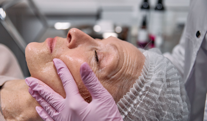
    A board-certified dermatologist conducting a professional medical skin evaluation on an adult patient's face at Chattanooga Skin and Cancer Clinic.
    Board-certified providers offer clinical evaluations for a wide range of chronic and acute skin conditions in Southeast Tennessee.

    Most people make their first dermatology appointment for one of two reasons: a suspicious spot they have been watching, or acne that will not clear up no matter what they try. Both are completely valid reasons to come in. The part that surprises most new patients is what they learn once they are there.

    Dermatology covers a wide range of conditions affecting the skin, hair, and nails. Skin cancer screenings and acne treatment are part of it, but so are chronic inflammatory conditions, hair loss, nail disorders, rashes that have defied every over-the-counter remedy, and conditions that started in childhood and followed a person into adulthood. Dermatologists are medical specialists with years of training specifically in skin health, which means the scope of what they diagnose and treat is considerably broader than most patients expect.

    At Chattanooga Skin and Cancer Clinic, patients have been coming in since 1973 for exactly that reason. Some know what they need. Others arrive with a condition their primary care doctor referred them for, or a symptom they have been trying to manage on their own. This post walks through what dermatologists treat, why some conditions belong in a specialist’s office rather than a general practitioner’s, and what patients in the Chattanooga area can expect when they come in.


    What Is a Dermatologist and How Is Their Training Different?

    A dermatologist is a physician who completed medical school, then spent three or more additional years in a dermatology residency focused entirely on skin, hair, and nail conditions. That residency includes surgical training, dermatopathology, and exposure to thousands of conditions ranging from common to rare. Board certification follows a rigorous examination process.

    This level of specialization matters because the skin is the body’s largest organ, and its conditions are numerous. More than 3,000 distinct skin conditions exist, according to the American Academy of Dermatology. A primary care physician can identify and treat many of the most common ones, but conditions that are chronic, resistant to standard treatment, or require a visual and clinical expertise built through years of practice are best managed by a specialist.

    All providers at Chattanooga Skin and Cancer Clinic are board-certified, and the practice has been operating in Southeast Tennessee for over 50 years. That depth of experience shapes how providers approach everything from a first-time skin check to a complex surgical case.


    Does a Dermatologist Only Treat Skin Cancer?

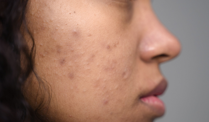
    Close-up of a patient with stubborn acne blemishes on their cheek seeking medical treatment from a dermatologist in Cleveland, TN.
    Persistent acne, driven by hormonal shifts or genetics, often requires a personalized prescription approach from a specialist.

    Skin cancer is one of the most important things a dermatologist diagnoses and treats, but it is far from the only one. The misconception that dermatology is primarily about cancer screenings keeps a lot of people from seeking care for conditions that are making their daily lives harder.

    Skin cancer detection and treatment, including Mohs micrographic surgery for basal cell and squamous cell carcinomas, is a core part of what the team at Chattanooga Skin and Cancer Clinic does. Mohs surgery is available at the Chattanooga and Cleveland locations and offers the highest cure rate for eligible skin cancers while preserving as much healthy tissue as possible. Melanoma screening, diagnosis, and treatment are also part of the practice’s focus.

    Outside of cancer, dermatologists manage a full range of medical conditions. Chronic inflammatory skin diseases like psoriasis, eczema, and rosacea require ongoing management that goes well past what an over-the-counter cream can provide. Acne, particularly adult acne driven by hormonal shifts, often needs a prescription approach that only a provider can prescribe. Rashes, allergic reactions, infections, warts, cysts, and moles all fall within a dermatologist’s scope of practice. So do hair loss, nail disorders, and conditions in children that can start as early as infancy.


    What Chronic Skin Conditions Can a Dermatologist Help Manage?

    Chronic conditions are where dermatology makes some of its most meaningful impact. These are conditions that do not go away on their own and that get worse without proper management. Living with one of them long-term without specialist care often means cycles of flare-ups, missed work, disrupted sleep, and real psychological strain.

    Is Psoriasis Something a Dermatologist Treats?

    A female dermatologist in a white coat explaining prescription biologics and targeted treatments to a patient in Chattanooga, Tennessee.
    Modern medical dermatology utilizes advanced targeted therapies and biologics to manage severe chronic skin conditions effectively.

    Psoriasis is an autoimmune condition that causes the skin to produce cells too rapidly, resulting in thick, scaly plaques that are often red, itchy, and uncomfortable. It shows up most often on the elbows, knees, scalp, and lower back, but it can appear anywhere on the body. Psoriasis is also associated with psoriatic arthritis, which affects joints and requires coordinated care.

    Treatment options have advanced considerably in recent years. Biologics and targeted therapies have given patients with moderate to severe psoriasis access to outcomes that were not possible even a decade ago. A dermatologist stays current on those options and can match the right treatment to the patient’s specific disease pattern, severity, and medical history.

    Can a Dermatologist Help With Eczema?

    Eczema, or atopic dermatitis, is one of the most common skin conditions in the United States, affecting about 31 million Americans according to the National Eczema Association. It causes dry, inflamed, itchy skin that can crack, bleed, and become infected when scratched. In children, it often appears on the face and in the skin creases. Adults tend to see it on the hands, neck, and inside of the elbows.

    Managing eczema well requires understanding a patient’s triggers, their skin barrier function, and the appropriate prescription options for their age and severity. The team at Chattanooga Skin and Cancer Clinic sees pediatric patients from infancy through adolescence and adult patients at all three locations. Consistent dermatology care for eczema reduces the frequency and severity of flares in a meaningful way.

    What About Rosacea?

    Rosacea is a chronic inflammatory condition that causes persistent facial redness, visible blood vessels, and sometimes acne-like bumps. It affects the cheeks, nose, forehead, and chin most often, and it is frequently mistaken for flushing or sensitive skin. Without treatment, rosacea progresses over time.

    Common triggers include heat, sun exposure, alcohol, spicy food, and stress. A dermatologist can confirm the diagnosis, identify a patient’s specific subtype, and recommend a combination of topical treatments, oral medications, and trigger management strategies. For patients in Chattanooga and the surrounding area, getting a definitive diagnosis from a board-certified provider is the starting point for effective long-term control.


    What Acute Conditions Bring Patients to a Dermatologist?

    Not every dermatology visit is about a chronic condition. Plenty of patients come in for something that developed recently and needs a prompt evaluation.

    Mole removal and evaluation is one of the most common acute reasons for a dermatology visit. A mole that has changed in size, shape, or color, or one that bleeds or itches without explanation, needs to be looked at by a trained eye. A dermatologist can examine the mole with a dermatoscope, determine whether a biopsy is necessary, and walk the patient through the results.

    Warts, cysts, and skin tags are also common reasons patients come in. These are benign growths, but they can be cosmetically bothersome, uncomfortable, or positioned in a way that causes irritation. Treatment options range from freezing to excision depending on the type and location. Rashes, allergic reactions, and skin infections round out the acute side of dermatology practice. Many patients come in after a rash has persisted for weeks without improvement, or after an over-the-counter treatment has made things worse rather than better. A provider can identify the underlying cause and prescribe the right course of action.


    Do Dermatologists Treat Children?

    Yes, and pediatric dermatology is an area where specialist care makes a significant difference. Children experience many of the same conditions as adults, but the presentation can differ, and treatment protocols are adjusted for age and developmental stage.

    Eczema is one of the most common pediatric skin conditions and one of the most underdiagnosed. Infants can develop it within the first few months of life. Warts, molluscum contagiosum, birthmarks, and scalp conditions are also common in children and teenagers. Acne in adolescents is another major area where a dermatologist provides real value, particularly when over-the-counter products have not produced results and the condition is starting to affect a young person’s confidence.

    At Chattanooga Skin and Cancer Clinic, pediatric and adolescent patients are seen at the Chattanooga and Cleveland locations. The practice has served families across the region for generations, which means a parent who came in as a teenager may now be bringing their own child in for care.


    When Should You See a Dermatologist Instead of Your Primary Care Doctor?

    Primary care physicians handle a wide range of skin issues well, and many straightforward cases can be managed at that level. A dermatologist is the right call when a condition is not responding to standard treatment, when it keeps coming back, when a visual evaluation from a trained specialist is needed, or when the patient wants access to the full range of treatment options including prescription biologics, surgical procedures, and advanced diagnostic tools.

    Annual full-body skin checks are something dermatologists do that general practitioners rarely perform in the same depth. A 15-minute full-body exam by a board-certified dermatologist covers areas a patient cannot easily see on their own, and it creates a documented baseline that makes future changes easier to identify. For patients over 35, for those with a family history of skin cancer, and for anyone who has spent significant time in the sun over the years, that annual check is worth making part of the regular health routine.

    Chattanooga Skin and Cancer Clinic accepts new patients at all three locations, in Chattanooga, Cleveland, and Kimball. No referral is required to schedule an appointment.


    Frequently Asked Questions

    Do I need a referral to see a dermatologist at Chattanooga Skin and Cancer Clinic? No. Chattanooga Skin and Cancer Clinic accepts new patients directly at all three locations without a referral. Appointments can be scheduled by calling the location nearest to you or visiting chattskinandcancer.com.

    What should I bring to my first dermatology appointment? Bring a list of your current medications and any supplements you take. Write down your skin concerns before the appointment, including how long each one has been present and what you have already tried. If you have a spot that comes and goes, take a photo of it when it is active. Coming with clean skin, free of heavy makeup or lotion in the areas you want evaluated, also helps the provider get the clearest view possible.

    Can a dermatologist help with hair loss? Yes. Hair loss has many causes, including autoimmune conditions, hormonal changes, nutritional deficiencies, and inflammatory scalp diseases. A dermatologist can evaluate the pattern and characteristics of hair loss, run relevant testing, and recommend treatment options. The scalp is skin, and scalp health is within a dermatologist’s scope of practice.

    How often should someone get a full-body skin check? For most adults, once a year is the standard recommendation. Patients with a personal or family history of skin cancer, a large number of moles, or significant past sun exposure may benefit from more frequent monitoring. Your provider at Chattanooga Skin and Cancer Clinic can recommend the right schedule based on your individual history during your first visit.

    Does Chattanooga Skin and Cancer Clinic see patients of all ages? Yes. The practice treats patients from infancy through elderly, making it a resource for the entire family. Pediatric and adolescent patients are seen at the Chattanooga and Cleveland locations. The Kimball location serves adult patients and handles medical dermatology conditions.

    What is the difference between medical and cosmetic dermatology? Medical dermatology focuses on diagnosing and treating conditions that affect health, including skin cancer, chronic inflammatory diseases, infections, and hair and nail disorders. Cosmetic dermatology focuses on aesthetic procedures like injectables and resurfacing treatments. Chattanooga Skin and Cancer Clinic is a medical dermatology practice. The providers focus on health-based care rather than cosmetic services.

  • Is That Spot on Your Skin Normal? 6 Warning Signs You Shouldn’t Ignore

    Is That Spot on Your Skin Normal? 6 Warning Signs You Shouldn’t Ignore


    Is That Spot on Your Skin Normal? 6 Warning Signs You Shouldn’t Ignore

    You notice something on your arm that wasn’t there before. It’s small, maybe slightly raised, and it looks a little different from the freckles you’ve had for years. You tell yourself it’s probably nothing. A few weeks pass, and it’s still there, maybe a touch bigger. That moment of uncertainty is exactly when a dermatologist visit makes a real difference. Skin changes are easy to brush off, but knowing which ones deserve a closer look can protect your health in a meaningful way.

    Skin cancer is the most commonly diagnosed cancer in the United States. The American Cancer Society estimates that over 100,000 new cases of melanoma alone are diagnosed each year. The good news is that most skin cancers, when caught early, are highly treatable. The tricky part is knowing what to watch for. Not every spot or mark is cause for alarm, but certain changes to your skin are clear signals to get checked by a board-certified dermatologist. Here are six warning signs that should never be ignored.


    Is a Spot Changing in Size, Shape, or Color?

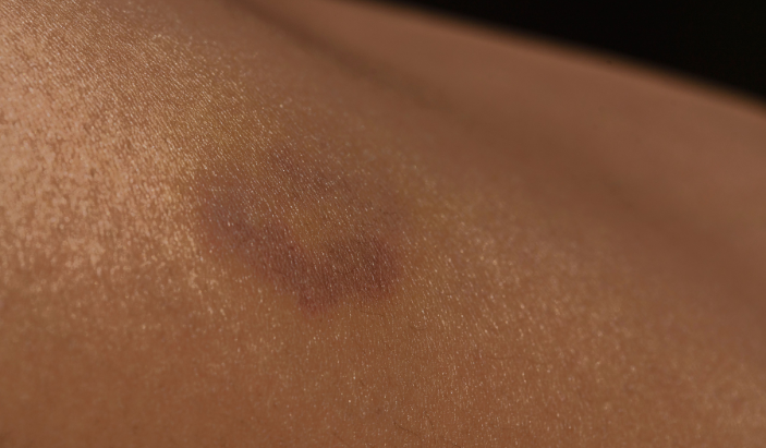
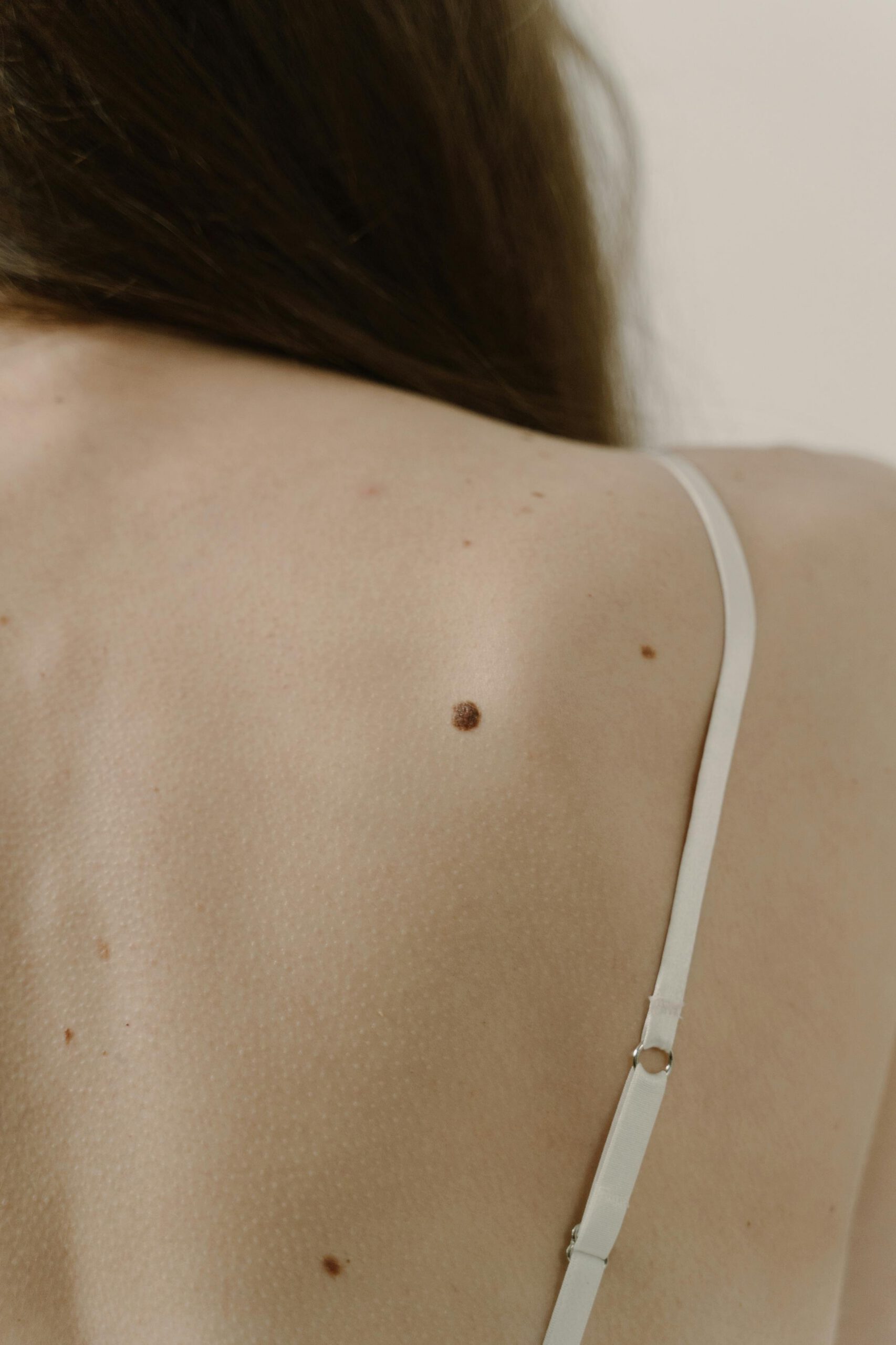
    Close-up of an irregular dark skin discoloration or asymmetrical mole needing evaluation by a Cleveland TN dermatologist.
    A dark, changing, or asymmetrical skin spot is a clear warning sign that warrants a professional evaluation.

    Change is the single most important word in skin cancer detection. A mole or spot that has looked the same for years and suddenly starts to shift is worth taking seriously. Board-certified dermatologists use the ABCDE rule to evaluate these changes. A stands for Asymmetry, meaning one half of a spot doesn’t match the other. B is for Border irregularity, where edges appear ragged, notched, or blurred. C covers Color variation, where a single spot contains multiple shades of brown, black, red, or white. D refers to Diameter, specifically any growth larger than six millimeters, roughly the size of a pencil eraser. E is for Evolution, the catch-all for any noticeable change over time.

    Any spot that checks more than one of those boxes warrants a professional skin exam. At Chattanooga Skin and Cancer Clinic, full-body skin checks are part of routine care, and the team has experience identifying changes that patients commonly overlook on their own.


    Does the Spot Bleed Without Any Obvious Cause?

    Spontaneous bleeding from a mole, lesion, or skin growth is a symptom that demands prompt attention. Healthy skin does not bleed on its own. If you notice a spot that bleeds after minimal contact, such as light rubbing from clothing or gentle washing, that behavior is abnormal and should be evaluated quickly.

    Basal cell carcinoma, one of the most common forms of skin cancer, can appear as a pearly or flesh-colored bump that bleeds easily when bumped or scratched. Squamous cell carcinoma sometimes presents similarly. Neither type is automatically dangerous when caught early, but both require professional diagnosis and treatment. Waiting on a bleeding spot is a risk not worth taking.


    Is There a Sore That Won’t Heal?

    A cut or scrape that doesn’t heal within two to four weeks is a red flag. Most minor skin wounds close up fairly quickly under normal circumstances. When a sore persists, reopens, or crusts over repeatedly without actually healing, that pattern can point to an underlying issue, including early skin cancer.

    Basal cell carcinomas sometimes look like open sores that appear to heal and then return. They can also resemble a flat, scar-like lesion with a pale or slightly yellow tint. If you have a wound that has lingered for more than a month without clear improvement, a dermatology appointment is the right next step.


    Has a Mole Started to Look Different from the Others?

    Dermatologists sometimes refer to this as the “ugly duckling” sign. Most people have moles that share a general family resemblance in terms of size, shape, and color. A mole that stands out as visibly different from the rest deserves attention. It doesn’t need to look dramatic to be suspicious. Sometimes the difference is subtle: a single mole that is darker, larger, or more irregular than everything else on your skin.

    This is one reason annual skin checks with a board-certified dermatologist are so useful. A trained eye can scan your full body and identify outliers that you simply cannot see yourself, especially on your back, scalp, and behind the ears. These are areas where many skin cancers develop precisely because they go unnoticed.


    Do You Notice a Pink, Pearly, or Waxy Growth?

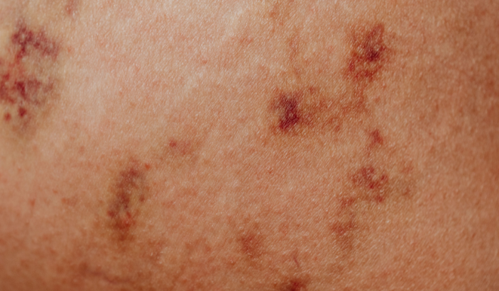
    Close-up of sun-damaged skin with multiple red and brown spots requiring a professional skin check in Kimball TN.
    Multiple clustered spots or scaly, red patches should be evaluated for squamous or basal cell carcinoma.

    Not all skin cancers look like dark, irregular moles. Basal cell carcinoma, the most common type, often presents as a small, shiny bump with a pink or translucent quality, sometimes with visible blood vessels near the surface. It can also appear as a flat, brownish scar-like patch. These subtle presentations are easy to dismiss as a harmless skin tag or minor blemish.

    Squamous cell carcinoma can show up as a rough, scaly patch of red skin, a firm bump, or a raised growth with a depressed center. Both cancers are highly treatable with early intervention. Mohs micrographic surgery, available at Chattanooga Skin and Cancer Clinic’s Chattanooga and Cleveland locations, is the most precise treatment option for these cancers and delivers cure rates above 99 percent for appropriate candidates.


    Is There Persistent Itching, Tenderness, or Unusual Sensation in a Spot?

    Skin spots that itch, burn, or feel tender without an obvious cause are worth noting. Occasional itchiness from dry skin is completely normal. A specific spot that consistently draws your attention because of sensation, especially combined with any visible change, is a different matter.

    Melanoma, the most serious form of skin cancer, can sometimes cause itching or tenderness in an affected mole. These sensations don’t always accompany skin cancer, but when they pair with other warning signs, they strengthen the case for getting a professional evaluation.


    When Should You Schedule a Skin Cancer Screening?

    A woman examining her face in a bathroom mirror for skin changes before scheduling a skin cancer screening in Chattanooga.
    Monitoring your skin at home is a vital first step in early skin cancer detection.

    If you spot any of the six warning signs above, schedule an appointment with a dermatologist as soon as possible. You don’t need to wait until your annual checkup if something looks or feels off. Early action consistently leads to better outcomes across every type of skin cancer.

    Beyond responding to specific concerns, adults with a family history of skin cancer, a personal history of frequent sun exposure, or a record of sunburns should schedule annual full-body skin checks. People with lighter skin tones or a high number of moles carry a higher baseline risk. Southeast Tennessee’s sunny climate means residents in Chattanooga, Cleveland, and Kimball get significant UV exposure throughout the year, which adds up over time.

    Chattanooga Skin and Cancer Clinic has served patients across the Tennessee Valley since 1973. All providers are board-certified dermatologists with deep experience in skin cancer detection, diagnosis, and treatment. Three convenient locations across Chattanooga, Cleveland, and Kimball make it straightforward to get the care you need close to home.


    Frequently Asked Questions

    How do I know if a mole is dangerous?

    Use the ABCDE rule as a starting point: Asymmetry, Border irregularity, Color variation, Diameter over six millimeters, and Evolution over time. Any mole that meets one or more of those criteria should be evaluated by a board-certified dermatologist. A professional skin check is the only reliable way to confirm whether a spot is benign or needs further attention.

    Can skin cancer appear somewhere other than a mole?

    Yes. Skin cancer can develop on any part of the body, including areas with no prior moles. Basal cell and squamous cell carcinomas often appear as growths, patches, or sores that don’t look like traditional moles at all. Some skin cancers develop on the scalp, ears, lips, palms, soles of the feet, and even under the nails.

    Is it normal for a mole to itch occasionally?

    Occasional mild itching in a mole can happen without any serious cause. Persistent itching, especially combined with visible changes such as growth, color shift, or irregular borders, is a reason to get checked. Itching alone is not a definitive sign of cancer, but it becomes more meaningful when it accompanies other warning signs.

    What happens during a full-body skin cancer screening?

    A full-body skin check is a head-to-toe visual examination performed by a dermatologist. The provider examines every area of your skin, including the scalp, back, and areas between the toes. The appointment takes about 15 to 30 minutes. If the dermatologist finds anything suspicious, they may perform a biopsy that same visit or schedule a follow-up. The exam is painless and doesn’t require any special preparation beyond removing clothing.

    How often should I see a dermatologist for skin cancer screening?

    Most adults benefit from an annual full-body skin check. People with a personal or family history of skin cancer, a high number of moles, or significant past sun exposure should discuss a more frequent schedule with their dermatologist. The right frequency depends on your individual risk profile.

    What is Mohs surgery and who is it for?

    Mohs micrographic surgery is a precise, layer-by-layer technique for removing basal cell and squamous cell carcinomas. It preserves the maximum amount of healthy tissue while achieving the highest cure rates available for these cancers. Mohs is particularly effective for tumors on the face, ears, hands, or feet where tissue conservation matters. The procedure is performed in a single visit. Chattanooga Skin and Cancer Clinic offers Mohs surgery at both the Chattanooga and Cleveland locations.


    Ready to Get Your Skin Checked?

    Catching something early is the most effective thing you can do for your skin health. If you’ve noticed any of the warning signs above, or if it’s simply been a while since your last professional skin exam, the team at Chattanooga Skin and Cancer Clinic is ready to help.

    Appointments are available at three locations across Southeast Tennessee:

    Chattanooga: 6061 Shallowford Road, Chattanooga, TN 37421 | 423-899-2700

    Cleveland: 3891 Adkisson Drive, Cleveland, TN 37312 | 423-479-8648

    Kimball: 400 Dixie Lee Center Rd Ste. C, Kimball, TN 37347 | 423-815-9975

    All three offices are open Monday through Friday. Visit chattskinandcancer.com to request an appointment online.

  • The 4 Types of Rosacea Explained: Which Type Do You Have?

    The 4 Types of Rosacea Explained: Which Type Do You Have?

    The 4 Types of Rosacea Explained: Which Type Do You Have?

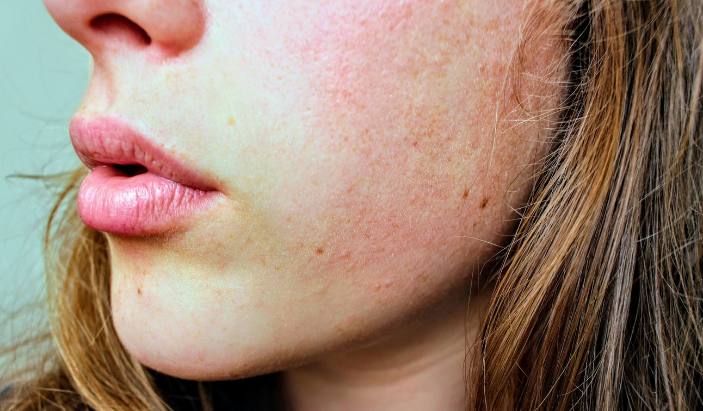
    Close-up of a patient's cheek with hyperpigmentation and sun damage, showcasing skin rejuvenation concerns
    Spotting the early signs of sun damage or uneven texture? Our Kimball skincare experts can help restore your skin’s natural glow.

    Rosacea is one of the most misunderstood skin conditions out there. A lot of people spend years treating what they think is adult acne, dry skin, or sun damage before a dermatologist finally connects the dots. The redness keeps coming back. The skin feels sensitive no matter what products you use. Something is clearly going on, but the label stays elusive. If that sounds familiar, there is a good chance rosacea is the reason.

    What makes rosacea especially tricky is that it does not look the same on every person. Some people develop flushing and broken blood vessels. Others deal with acne-like breakouts. Some have thickened skin, and others struggle with eye irritation that they would never connect to a skin condition. Rosacea has four distinct subtypes, and understanding which one you have is the starting point for getting it properly managed.

    According to the National Rosacea Society, more than 16 million Americans are affected by rosacea, and many of them do not realize they have it. This article breaks down each of the four types, what they look like, how they differ, and what you can do about them.


    What Is Rosacea and Why Does It Come in Different Types?

    Rosacea is a chronic skin condition that causes redness, inflammation, and a range of other symptoms primarily on the face. It tends to cycle through flares and calmer periods, often triggered by heat, sun, spicy food, alcohol, stress, or certain skincare products. There is no cure, but with the right treatment plan, most people can keep it well under control.

    The reason rosacea has subtypes is that it affects different structures of the skin in different ways. The blood vessels near the surface, the oil glands, the connective tissue, and even the eyes can all be involved depending on the person. Doctors and researchers use a subtype classification system to describe these patterns and match them to the most effective treatments.

    It is worth knowing that subtypes are not always mutually exclusive. Some people experience two or more types at once, or see one subtype evolve into another over time if the condition goes untreated. That is another reason early diagnosis from a board-certified dermatologist matters so much.


    What Is Subtype 1 and How Does Erythematotelangiectatic Rosacea Show Up?

    Subtype 1 is called erythematotelangiectatic rosacea, often abbreviated as ETR. The name is a mouthful, but the presentation is fairly straightforward: persistent redness across the cheeks, nose, chin, or forehead, often accompanied by flushing and visible blood vessels close to the skin’s surface.

    People with ETR frequently report that their face feels like it is burning or stinging, even without an obvious trigger. The skin tends to be sensitive and reactive, flaring up in response to temperature changes, exercise, wind, hot beverages, or sun exposure. The redness does not fade the way a normal flush would. It lingers, and over time, the tiny blood vessels known as telangiectasias become more visible, creating a web of fine red or pink lines across the face.

    This subtype is the most commonly recognized form of rosacea. Many people with ETR try to treat the redness with moisturizers or over-the-counter products, but those rarely address what is actually happening beneath the surface. A dermatologist can prescribe topical medications that reduce redness or recommend in-office laser and light treatments that target the visible blood vessels directly.


    What Is Subtype 2 and How Is Papulopustular Rosacea Different From Acne?

    Young male patient experiencing facial redness and rosacea-like flushing on the cheeks, illustrating sensitive skin treatments.
    Persistent facial flushing or redness can be managed with a custom treatment plan at our Kimball clinic.

    Subtype 2 is papulopustular rosacea, and it is the one most frequently mistaken for acne. It causes red bumps called papules and pus-filled blemishes called pustules, usually concentrated in the central face. That combination of breakouts plus background redness is the classic picture of this subtype.

    The key difference between papulopustular rosacea and acne comes down to what you do not see. Rosacea does not produce blackheads or whiteheads. Acne does. If you have what looks like adult acne but there are no blackheads in the mix, rosacea becomes a much more likely explanation. Age is another clue. Papulopustular rosacea is more common in adults, particularly women in their 30s, 40s, and 50s.

    Using standard acne treatments on this subtype can backfire badly. Many acne products contain ingredients like benzoyl peroxide or retinoids that are simply too harsh for rosacea-prone skin. They strip and irritate the skin barrier, making the redness and breakouts worse. A proper diagnosis is the only way to avoid that cycle. A dermatologist can confirm which condition you are dealing with and prescribe treatments like topical azelaic acid, metronidazole, or oral antibiotics that are designed for rosacea specifically.


    What Is Subtype 3 and Who Does Phymatous Rosacea Typically Affect?

    Subtype 3 is phymatous rosacea, the rarest and most dramatic presentation of the condition. It causes a thickening and irregular texture of the skin due to the enlargement of oil glands and a buildup of connective tissue. The nose is the most commonly affected area, and when it progresses significantly, the condition is called rhinophyma.

    Rhinophyma creates a bulbous, bumpy appearance on the nose, with enlarged pores and a rough texture. It can also affect the chin, forehead, ears, and eyelids, though this is less frequent. Phymatous rosacea is much more common in men than in women, and the reasons for that are not entirely clear, though hormonal factors are thought to play a role.

    This subtype tends to develop gradually over years, often in people who had untreated rosacea for a long time. That makes it one of the stronger arguments for getting rosacea evaluated and managed early. Once phymatous changes develop, they do not reverse on their own. Treatment in the earlier stages involves prescription medications to slow progression. More advanced cases may require laser resurfacing or surgical intervention to reshape the tissue.


    What Is Subtype 4 and How Does Ocular Rosacea Affect the Eyes?

    Subtype 4 is ocular rosacea, and it is the most overlooked type because many people do not connect eye symptoms to a skin condition. Ocular rosacea affects the eyes and the skin around them, causing redness, irritation, dryness, and a persistent gritty or burning sensation. The eyelids may become swollen, crusty, or inflamed, a condition called blepharitis.

    Some people with ocular rosacea develop it alongside one of the other subtypes. Others experience the eye symptoms before any skin changes appear, which makes diagnosis particularly confusing. Left unmanaged, ocular rosacea can lead to sensitivity to light, blurred vision, and in more serious cases, corneal damage.

    Treatment for ocular rosacea often involves a combination of approaches: warm compresses, lid hygiene routines, prescription eye drops, and in some cases oral antibiotics that reduce inflammation. A dermatologist and an ophthalmologist sometimes work together to manage this subtype, especially when both skin and eye symptoms are present.


    How Do You Know Which Type of Rosacea You Have?

    Identifying your subtype is not something you can do reliably on your own, and trying to piece it together from an internet search can lead you in the wrong direction. A board-certified dermatologist evaluates your skin in person, considers your symptom history, and identifies not just which subtype is present but whether multiple subtypes are overlapping.

    The distinction matters because treatment varies meaningfully by subtype. The approach for ETR focuses on reducing vascular reactivity and protecting a sensitive skin barrier. Papulopustular rosacea calls for anti-inflammatory treatments. Phymatous changes may require procedures. Ocular involvement often needs specialized eye care in addition to dermatology management. A one-size approach simply does not work across all four.

    At Chattanooga Skin and Cancer Clinic, board-certified dermatologists have been evaluating and treating rosacea patients across the Chattanooga, Cleveland, and Kimball area since 1973. With three convenient locations and same-team continuity of care, patients get a thorough assessment and a treatment plan that fits their specific presentation.


    What Triggers Make Rosacea Worse Across All Four Types?

    Regardless of subtype, rosacea responds to a similar set of triggers. Sun exposure is one of the most common, and Tennessee summers can be particularly rough on rosacea-prone skin. UV radiation dilates blood vessels and causes lasting inflammation over time. Daily broad-spectrum sunscreen is a non-negotiable part of managing any type of rosacea.

    Heat, spicy foods, alcohol (particularly red wine), hot beverages, stress, and intense exercise all commonly provoke flares. So do certain skincare ingredients: fragrances, alcohol-based toners, witch hazel, and harsh exfoliants. Keeping a simple trigger diary helps identify personal patterns, since not everyone reacts to the same things.

    The goal of trigger management is not to eliminate every possible irritant from your life. It is to identify your top offenders so you can minimize them where it makes a real difference. Combined with prescription treatment from a dermatologist, trigger awareness gives patients the best shot at keeping rosacea calm long-term.


    Frequently Asked Questions About the Types of Rosacea

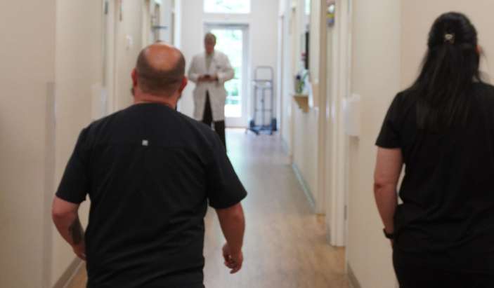
    Medical staff and dermatologist walking down the hallway of Chattanooga Skin and Cancer Clinic, a premier dermatology clinic.
    Our expert dermatology team in Kimball working behind the scenes to bring you personalized, medical-grade skin solutions

    Can you have more than one type of rosacea at the same time?

    Yes. It is fairly common for people to experience two subtypes simultaneously. Subtype 1 and Subtype 2 often occur together, with persistent redness and visible blood vessels accompanying acne-like breakouts. Ocular symptoms can also appear alongside any of the skin-based subtypes.

    Does rosacea subtype change over time?

    It can. Rosacea is a progressive condition in many people, meaning it tends to worsen if left untreated. Subtype 1 can evolve to include the papules and pustules of Subtype 2 over time. Phymatous changes typically develop after years of chronic, unmanaged inflammation. Getting diagnosed and starting treatment early is the best way to slow or prevent that progression.

    Is rosacea more common in certain skin tones?

    Rosacea is most frequently diagnosed in people with fair skin, but it affects people of all skin tones. On medium to deeper skin tones, it often goes unrecognized because the classic redness is harder to see. Symptoms like burning, stinging, bumps, or eye irritation may still be present, and a dermatologist familiar with rosacea presentations across diverse skin tones can make an accurate diagnosis.

    Are there rosacea treatments that work for all four types?

    Some treatments overlap across subtypes, particularly topical and oral anti-inflammatory medications. That said, the most effective treatment plans are specific to the subtype. Laser treatments that target blood vessels are most useful for Subtype 1. Antibiotic therapies are more central to Subtype 2. Subtype 3 may require procedural intervention. Subtype 4 often needs targeted eye care. Treatment works best when it is matched to what is actually happening in your skin.

    When should you see a dermatologist about rosacea?

    As soon as you suspect it. Rosacea does not improve on its own, and the longer it goes unmanaged, the more ingrained the changes to the skin can become. If you have persistent facial redness, recurring breakouts without blackheads, skin that feels easily irritated, or any eye discomfort that your eye doctor has not fully explained, a dermatology evaluation is the right move. Early treatment produces the best long-term results.


    Ready to Get a Clear Answer About Your Skin?

    Living with rosacea is manageable, but only once you understand what you are actually dealing with. Guessing at a diagnosis and cycling through the wrong products wastes time and often makes the condition worse. The four subtypes of rosacea are distinct enough that proper identification genuinely changes the treatment path.

    The team at Chattanooga Skin and Cancer Clinic includes board-certified dermatologists who have spent decades treating rosacea patients across Southeast Tennessee. Appointments are available in Chattanooga, Cleveland, and Kimball. Call the location nearest you or request an appointment online at chattskinandcancer.com.

  • Eczema in Babies and Toddlers: What Parents Need to Know

    Eczema in Babies and Toddlers: What Parents Need to Know


    Your baby wakes up again at 2 a.m., scratching at the same raw patches of skin on their cheeks and elbows. You’ve tried switching detergents, avoiding certain foods, and slathering on whatever lotion was at the top of the search results. Nothing is really working, and the pediatrician used a word you’ve been reading about ever since: eczema. If that scenario sounds familiar, you are not alone. Eczema is one of the most common skin conditions in young children, and it is also one of the most misunderstood.

    Eczema in babies and toddlers affects roughly 10 to 20 percent of children in the United States, according to the National Eczema Association. It tends to show up in the first year of life, often before a baby’s first birthday. For many parents, the diagnosis raises more questions than answers. What exactly is eczema? What causes it to flare? How do you manage it day to day, and when does a child need to see a dermatologist? This article answers all of those questions clearly and practically, so parents in the Chattanooga area and beyond can feel confident taking care of their child’s skin.


    What Is Eczema and Why Does It Affect So Many Babies?

    A crying infant showing signs of facial discomfort and skin sensitivity from baby eczema.
    Severe itching and skin discomfort from infant eczema can deeply disrupt a baby’s comfort and sleep.

    Eczema, medically known as atopic dermatitis, is a chronic inflammatory skin condition where the skin’s protective barrier does not function properly. A healthy skin barrier locks in moisture and keeps irritants and allergens out. In children with eczema, that barrier is compromised, meaning the skin loses water more easily and becomes vulnerable to environmental triggers.

    The result is skin that is dry, itchy, and prone to red, inflamed patches. In babies, these patches often appear on the face, especially the cheeks and forehead. As children grow into toddlers, eczema tends to shift to the elbow creases, behind the knees, and around the wrists. The itching can be intense, and because babies and toddlers cannot stop themselves from scratching, the skin can break open, which raises the risk of infection.

    Eczema is not contagious. It is not caused by poor hygiene or anything a parent did wrong. Genetics play a large role. Children with a parent or sibling who has eczema, asthma, or seasonal allergies are significantly more likely to develop it themselves. This connection, called the atopic march, means that early eczema in infants can sometimes be a predictor of later allergies or asthma.


    How Do You Know If Your Baby Has Eczema?

    Eczema can look different depending on the child’s age and skin tone. In lighter skin, inflamed patches appear red or pink. In deeper skin tones, eczema may look more purple, brown, or grayish, which leads to it being missed or misdiagnosed more often in children of color.

    Common signs to watch for in babies under 12 months include dry, scaly patches on the cheeks, scalp, or forehead; oozing or crusting skin; and visible discomfort, especially when the baby is warm or after a bath. Toddlers often show eczema in the skin folds at the elbows, wrists, knees, and ankles. The skin may appear thickened or leathery in areas that have been scratched repeatedly over time.

    One hallmark of eczema is the cycle of flares and relative calm. A child might have clear skin for weeks, then break out after a trigger like cold weather, sweating, or exposure to a new product. That unpredictable pattern can make management feel frustrating, but understanding triggers is one of the most useful tools parents have.


    What Triggers Eczema Flares in Young Children?

    Triggers vary from child to child, but some of the most consistent culprits include dry air, sweat, synthetic fabrics, certain soaps and fragrances, dust mites, pet dander, and stress. In the Southeast, where Chattanooga’s climate includes both humid summers and cold winters, children with eczema can face year-round challenges as the environment shifts.

    Food allergies sometimes overlap with eczema, which leads many parents to assume that diet is the root cause. The relationship between eczema and food is real but complicated. The American Academy of Dermatology notes that food allergies are found in about 30 percent of children with moderate to severe eczema, with milk, eggs, wheat, soy, and peanuts being the most common culprits. Eliminating foods without guidance can be harmful to a young child’s nutrition. A board-certified dermatologist or allergist should evaluate whether food is genuinely contributing to a child’s flares before any dietary changes are made.

    Temperature is another major factor. Overheating causes sweating, which irritates the skin. Keeping bedrooms cool, dressing children in loose cotton layers, and avoiding heavy blankets at night can all reduce the frequency of flares.


    How Is Eczema in Babies and Toddlers Treated?

    There is no cure for eczema, but it is very manageable with a consistent routine. The foundation of eczema care for young children is moisturizing frequently and effectively. Applying a thick, fragrance-free cream or ointment immediately after a bath, while the skin is still slightly damp, helps lock in moisture before it evaporates. Creams and ointments outperform thin lotions for this purpose because they contain more lipid content and create a stronger barrier.

    Bathing in lukewarm water for five to ten minutes, then patting the skin dry before applying moisturizer, is a routine that dermatologists recommend consistently. Hot water strips the skin’s natural oils and worsens dryness. Fragrance-free, gentle cleansers should replace traditional soaps.

    When moisturizing alone does not control flares, a pediatric dermatologist may recommend topical corticosteroids. These prescription creams reduce inflammation and are safe when used correctly. Parents are often cautious about steroids, and that concern is understandable, but low-potency topical steroids used as directed on affected areas are a well-established treatment option. A dermatologist will choose the appropriate strength for the child’s age and the area being treated.

    Newer non-steroidal prescription treatments, such as topical calcineurin inhibitors, are also available for children who need ongoing management without repeated steroid use. For more severe cases, a pediatric dermatologist may discuss systemic treatments or biologic medications. Dupilumab received FDA approval for children as young as six months old in 2023, marking a meaningful step forward in treating moderate to severe eczema in infants.


    When Should You See a Dermatologist Instead of Waiting It Out?

    Pediatricians are a good starting point for mild eczema, but there are clear situations where a dermatologist’s expertise makes a real difference. A board-certified dermatologist has specialized training in skin conditions that goes beyond what most general practitioners receive.

    See a dermatologist if your child’s eczema is not improving after two to four weeks of consistent moisturizing and over-the-counter care. Seek care sooner if the skin appears infected, which may look like crusting, honey-colored discharge, increased redness, warmth, or swelling. Scratched eczema skin is an open door for bacterial infections, particularly staph, and those infections need prompt treatment.

    A dermatologist can also confirm the diagnosis when eczema looks similar to other conditions like psoriasis, seborrheic dermatitis, or contact dermatitis. Getting the right diagnosis matters because these conditions respond to different treatments. Treating one condition like another will not produce the results the child needs.

    Chattanooga Skin and Cancer Clinic has been caring for patients of all ages since 1973, including infants and toddlers with chronic skin conditions. All providers are board-certified dermatologists with experience in pediatric dermatology. With three locations across Chattanooga, Cleveland, and Kimball, families across Southeast Tennessee can access specialized care close to home.


    What Daily Habits Make the Biggest Difference for Kids with Eczema?

    A parent checking a baby's back for dry patches and toddler eczema flares in Chattanooga.
    Consistently checking your child’s skin folds and back helps track eczema triggers and flare-ups early.

    Consistency is everything with eczema management. Moisturizing once a day is helpful; moisturizing twice a day is better. The goal is to keep the skin barrier as intact as possible so it has less opportunity to react to triggers.

    Clothing choices matter more than most parents realize. Synthetic fabrics like polyester and nylon trap heat and create friction against the skin. Loose-fitting, 100 percent cotton clothing lets the skin breathe and reduces irritation. Wash new clothing before the first wear to remove manufacturing residues, and use fragrance-free, dye-free detergents consistently.

    Keeping a simple log of flares can help parents and dermatologists identify patterns. Note what the child ate, what products were used, changes in weather or environment, and how the skin responded. Over time, these records often reveal connections that are not obvious in the moment.

    Short, trimmed fingernails reduce the skin damage that comes from scratching during sleep. For infants who scratch at night, lightweight cotton mittens can protect the skin until they are old enough to understand not to scratch.


    Does Eczema in Babies Go Away on Its Own?

    Many children do experience significant improvement as they get older. Studies suggest that about 50 percent of children with infant eczema see it resolve or become much milder by age six. Another portion will continue to have flares into adolescence or adulthood, though often less severely than in early childhood.

    The trajectory is hard to predict for any individual child. Children with more severe early eczema, or those who also have food allergies or asthma, are more likely to have persistent symptoms. Early, consistent treatment and close management with a dermatologist give children the best chance at keeping flares minimal and protecting skin health long-term.

    Parents should also know that eczema does not reflect on their parenting. Many children with eczema go on to have completely normal, active childhoods when their condition is well managed.


    Frequently Asked Questions About Eczema in Babies and Toddlers

    Is eczema the same as dry skin?

    No. While dry skin is a feature of eczema, eczema is a chronic inflammatory condition with a genetic component. Simple dry skin responds to moisturizer and does not cause the chronic, itchy, inflamed patches that eczema does. If moisturizing alone is not controlling the problem, the skin condition likely needs a dermatologist’s assessment.

    Can breastfeeding or formula affect my baby’s eczema?

    There is some evidence that breastfeeding may offer a modest protective effect against developing eczema, but breastfeeding does not prevent it entirely and formula does not cause it. If a nursing mother suspects her diet is affecting her baby’s skin, speaking with a dermatologist or allergist is the right path forward rather than eliminating foods without guidance.

    Are topical steroids safe to use on a baby’s skin?

    Low-potency topical corticosteroids are considered safe for short-term use in infants and toddlers when prescribed by a dermatologist and applied as directed. Concerns about steroid side effects are most relevant when high-potency creams are overused for long periods. A board-certified dermatologist prescribes the appropriate strength and gives clear instructions on duration and frequency.

    What is the best moisturizer for baby eczema?

    Thick, fragrance-free creams or ointments tend to work better than thin lotions. Products like plain petroleum jelly, CeraVe Healing Ointment, or Vanicream Moisturizing Ointment are frequently recommended because they are fragrance-free and effective. The best moisturizer is one that is applied consistently and immediately after bathing while the skin is still slightly damp.

    When should I bring my toddler to Chattanooga Skin and Cancer Clinic for eczema?

    A toddler holding a stuffed animal while being examined at Chattanooga Skin and Cancer Clinic.
    Board-certified pediatric dermatologists offer advanced treatment plans for moderate to severe childhood eczema.

    Bring your child in if the eczema is not responding to at-home care after two to four weeks, if you see signs of infection, if the condition is affecting your child’s sleep or quality of life, or if you are unsure whether the diagnosis is correct. The practice’s board-certified dermatologists see patients across all age groups, including infants, at locations in Chattanooga, Cleveland, and Kimball. You can call the Chattanooga office at 423-899-2700 or the Cleveland office at 423-479-8648 to schedule an appointment.

    Getting Your Child the Right Care

    Eczema in babies and toddlers can feel relentless, especially in those early months when everything is new and sleep is already scarce. The good news is that with the right routine, the right triggers identified, and the right medical support in place, most children with eczema live comfortably and thrive.

    Chattanooga Skin and Cancer Clinic has been part of this community for more than 50 years, founded in 1973 and now serving patients at three locations across Southeast Tennessee. Every provider is board-certified, and the clinic treats patients from infancy through adulthood. If your baby or toddler is struggling with eczema and you are ready to move past guesswork, the team at Chattanooga Skin and Cancer Clinic is ready to help.

    Call 423-899-2700 for the Chattanooga location at 6061 Shallowford Road, 423-479-8648 for Cleveland at 3891 Adkisson Drive, or 423-815-9975 for the Kimball office at 400 Dixie Lee Center Rd. All three offices are open Monday through Friday, and new patient appointments are available.


  • Sunburn vs. Sun Poisoning: How to Tell the Difference and What to Do Next

    Sunburn vs. Sun Poisoning: How to Tell the Difference and What to Do Next


    You spent the day at the lake, maybe a little longer than planned. By evening, your skin is red and tender, and you feel run-down in a way that sleep alone does not seem to fix. Most people chalk that up to a bad sunburn and move on. Sometimes, though, what feels like a rough day in the sun is actually sun poisoning, a more serious reaction that calls for a different response.

    Sunburn and sun poisoning both result from overexposure to ultraviolet (UV) radiation. The difference comes down to severity and how your body reacts. Knowing which one you are dealing with helps you treat it properly and avoid complications that can turn a few uncomfortable days into a trip to urgent care or the dermatologist.


    What Is Sunburn, and Why Does It Happen?

    A woman with a severe sunburn on her shoulders sitting on a wooden bench by the water in Chattanooga, Tennessee.
    Spending a day out on the water can easily lead to severe UV damage and painful sunburns.

    Sunburn is the skin’s inflammatory response to UV radiation damage. When UV rays penetrate the skin, they damage the DNA in skin cells. The redness, warmth, and tenderness you feel are signs of that inflammation as your immune system responds to the injury.

    Symptoms of a typical sunburn include red or pink skin that is warm and sensitive to the touch, mild swelling, and peeling that begins a few days later as damaged skin cells shed. You may feel some fatigue and mild discomfort. These symptoms are generally confined to the skin itself. Most sunburns resolve on their own within three to five days with basic care.

    According to the Skin Cancer Foundation, a single blistering sunburn in childhood or adolescence more than doubles a person’s lifetime risk of developing melanoma. Even sunburns that do not blister accumulate damage over time. Repeated sunburns are one of the strongest preventable risk factors for skin cancer, which is why dermatologists take UV exposure so seriously.


    What Makes Sun Poisoning Different?

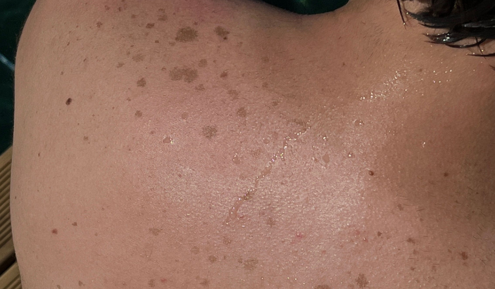
    Close-up of sun-damaged skin on a person's upper back with sun spots and freckles, highlighting the need for a dermatology screening in Kimball TN.
    Repeated UV exposure causes cumulative DNA damage, often resulting in sun spots, peeling, or changing moles.

    Sun poisoning is not a poisoning in the medical toxicology sense. The term refers to a severe sunburn that triggers a systemic reaction, meaning your body responds beyond the skin. Some people call it photodermatitis or polymorphous light eruption (PMLE), though those conditions involve specific immune reactions to sunlight and can differ from a straightforward overexposure reaction.

    The skin symptoms are more severe than a typical sunburn. Blistering, intense swelling, and skin that looks tight or leathery are common. The defining feature of sun poisoning, though, is that you feel sick. Nausea, vomiting, headache, dizziness, and fever separate sun poisoning from ordinary sunburn. Chills, dehydration, and in serious cases, a rapid heart rate or confusion can accompany these symptoms.

    Some people also develop a rash of small bumps or hives, which points toward an immune-mediated response to sun exposure. Fair-skinned individuals, people spending long hours outdoors at high altitudes or near reflective surfaces like water and snow, and those taking medications that increase sun sensitivity face higher risk.


    How Do You Tell the Two Apart?

    The clearest signal is how you feel overall. A sunburn hurts and looks bad, but you still feel relatively normal otherwise. Sun poisoning crosses into feeling genuinely ill. If you have nausea, a fever, dizziness, or a severe headache on top of your skin symptoms, sun poisoning is the more likely explanation.

    Look at the skin, too. Sunburn produces an even redness across the exposed area. Sun poisoning often brings blistering, significant swelling, or a rash pattern that looks unusual compared to regular redness. If the skin appears wet, oozing, or blistered across a large area of the body, that is a sign the reaction has gone beyond a mild sunburn.

    The timeline matters as well. Sunburn symptoms peak around 24 to 36 hours after exposure and then start to improve. Sun poisoning symptoms can intensify over the first day or two and take considerably longer to resolve. If things seem to be getting worse instead of better after 48 hours, that is worth paying attention to.


    What Should You Do If You Have a Sunburn?

    Treating a standard sunburn starts with cooling and hydration. Get out of the sun immediately and stay out. Cool (not cold) showers or compresses can reduce heat and discomfort in the skin. Drink extra water because UV radiation dehydrates the body even when you do not feel thirsty.

    Apply a fragrance-free moisturizer or aloe vera gel to the affected skin. Aloe has mild anti-inflammatory properties and can ease discomfort. Avoid products with added fragrances, alcohol, or numbing agents like benzocaine, which can irritate damaged skin or cause allergic reactions. Over-the-counter ibuprofen or naproxen helps reduce inflammation and pain and works better for sunburn than acetaminophen alone.

    Do not pop blisters if they form. Blisters are the skin’s natural protective barrier over the damaged area. Breaking them opens the wound to infection. If blisters do develop, keep the area clean and covered with a non-stick bandage. Once the skin starts peeling, let it happen on its own rather than pulling it off prematurely.


    What Should You Do If You Have Sun Poisoning?

    Sun poisoning with mild systemic symptoms, such as a slight headache and nausea, can often be managed at home with aggressive hydration, rest, and the same topical care used for sunburn. The key difference is that dehydration is a real risk. Water and electrolyte drinks are both important. If you cannot keep fluids down because of vomiting, that changes the situation.

    Seek medical attention if you have a fever above 103 degrees Fahrenheit, severe or spreading blisters, confusion or extreme dizziness, a rapid heartbeat, or if you are unable to stay hydrated due to persistent vomiting. Children, older adults, and anyone with a compromised immune system should seek care sooner rather than waiting to see if symptoms improve.

    Emergency care is appropriate for anyone showing signs of heat stroke alongside sun poisoning: confusion, loss of consciousness, no sweating despite extreme heat, or skin that looks gray or mottled. Heat stroke is life-threatening and requires immediate intervention.


    When Should You See a Dermatologist After Sun Exposure?

    A single bad sunburn may not require a dermatology visit, but several situations do. If you notice a rash that keeps returning with sun exposure, that points toward polymorphous light eruption or another photosensitivity condition, and a board-certified dermatologist can diagnose it and outline a management plan.

    Any new or changing spot on sun-damaged skin deserves a professional look. Repeated sun exposure increases the risk of basal cell carcinoma, squamous cell carcinoma, and melanoma, the three most common types of skin cancer. Catching these early makes treatment far more straightforward. Dermatologists recommend annual skin checks for anyone with a history of significant sun exposure, multiple sunburns, or a family history of skin cancer.

    If you live in the Chattanooga area, Chattanooga Skin and Cancer Clinic has provided medical dermatology care since 1973. The practice’s board-certified dermatologists see patients at three locations, including Chattanooga, Cleveland, and Kimball, and offer skin cancer screenings, mole evaluation, and treatment for chronic skin conditions.


    How Do You Protect Your Skin From UV Damage Going Forward?

    A close-up of a person applying sunscreen to a sun-exposed shoulder near the water, promoting skin cancer prevention.
    Applying broad-spectrum sunscreen is the most effective way to prevent both regular sunburn and systemic sun poisoning.

    The best defense against both sunburn and sun poisoning is consistent sun protection. Broad-spectrum sunscreen with SPF 30 or higher, applied 15 to 30 minutes before going outside and reapplied every two hours (or after swimming or sweating), forms the foundation of that protection. Most people apply far less sunscreen than needed for adequate coverage. Be generous on the face, neck, ears, and hands, which tend to accumulate the most UV exposure over a lifetime.

    UV-protective clothing, wide-brimmed hats, and sunglasses add another layer of defense, especially during peak UV hours between 10 a.m. and 4 p.m. Shade helps during those hours. Reflective surfaces like water, sand, and concrete intensify UV exposure even when cloud cover makes the day feel mild, so sun protection applies on overcast days as well.

    Certain medications, including some antibiotics, antihistamines, and diuretics, increase photosensitivity. If you take any prescriptions and plan to spend extended time outdoors, ask your doctor or pharmacist whether extra precautions make sense.


    Frequently Asked Questions About Sunburn and Sun Poisoning

    Can you get sun poisoning on a cloudy day?

    Yes. Up to 80 percent of UV rays pass through cloud cover. Overcast skies reduce but do not block UV radiation, and many serious sunburns and sun poisoning cases happen on cloudy days when people skip sunscreen because the sun does not feel intense.

    How long does sun poisoning last?

    Mild cases often resolve within two to three days. More severe cases with significant blistering or systemic symptoms can last up to a week or longer. If symptoms persist beyond a week or worsen after the first couple of days, see a doctor.

    Does tanning prevent sunburn?

    A tan offers minimal protection, roughly equivalent to an SPF of 2 to 4. It does not prevent UV damage. Each tanning session adds to cumulative DNA damage in the skin, which raises long-term skin cancer risk regardless of whether it results in a burn.

    Are some people more prone to sun poisoning?

    Fair-skinned individuals with less natural melanin face higher risk. People with a personal or family history of photosensitivity reactions, those at high altitudes, and anyone taking photosensitizing medications are also more susceptible. A history of previous sun poisoning episodes is one of the strongest predictors of future episodes.

    When does a sunburn require emergency care?

    Head to the emergency room or call 911 if you experience confusion, loss of consciousness, a rapid or irregular heartbeat, inability to keep fluids down, or signs of heat stroke alongside severe skin symptoms. These are medical emergencies that go beyond what at-home care can address.

    How does sun exposure contribute to skin cancer risk?

    UV radiation damages the DNA in skin cells, and that damage accumulates with each exposure. When DNA repair mechanisms cannot keep up, mutations can develop that lead to uncontrolled cell growth, which is how skin cancer forms. Melanoma, basal cell carcinoma, and squamous cell carcinoma are all strongly linked to UV exposure history.

    Should I schedule a skin check after a severe sunburn?

    A severe sunburn, especially one that blisters, is a reasonable prompt to schedule a baseline skin check with a dermatologist, particularly if you have other risk factors like fair skin, many moles, or a family history of skin cancer. Annual skin cancer screenings are a smart habit for anyone with a history of significant UV exposure.


    Sun damage adds up over a lifetime, and the choices you make now affect your skin health for years to come. If you are overdue for a skin check or have concerns about spots, moles, or a history of sunburns, Chattanooga Skin and Cancer Clinic is accepting new patients at all three locations. The clinic’s board-certified dermatologists specialize in medical dermatology and skin cancer detection, with more than 50 years of experience serving the greater Chattanooga region. Call 423-899-2700 to schedule in Chattanooga, 423-479-8648 in Cleveland, or 423-815-9975 in Kimball.

  • How Often Should You Get a Full-Body Skin Cancer Screening? (Age-by-Age Guide)

    How Often Should You Get a Full-Body Skin Cancer Screening? (Age-by-Age Guide)


    A full-body skin cancer screening is one of the most straightforward steps you can take to protect your health. A board-certified dermatologist checks your skin from head to toe, looking for moles, spots, or lesions that may need attention. The exam is quick, painless, and in many cases, it saves lives. Yet most people do not know how often they should go, or what the right schedule looks like for their age and risk level.

    Skin cancer is the most commonly diagnosed cancer in the United States, with the American Academy of Dermatology estimating that one in five Americans will develop it during their lifetime. Catching it early, while it is still confined to the surface of the skin, makes treatment far simpler and outcomes far better. That is why understanding the right screening schedule matters as much as the screening itself.


    What Does a Full-Body Skin Cancer Screening Actually Involve?

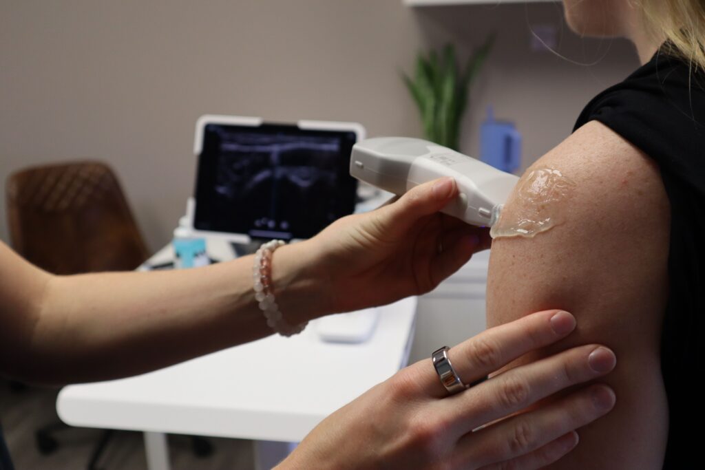
    A board-certified dermatologist at a Chattanooga skin clinic using an advanced screening device on a patient's upper arm.
    Modern dermatology clinics utilize precise tools and advanced techniques to identify and track suspicious spots early.

    A full-body skin cancer screening is a head-to-toe visual exam performed by a dermatologist. The doctor examines every area of your skin, including spots you cannot easily see yourself, such as your scalp, between your toes, behind your ears, and on your back. The appointment typically takes about 15 to 30 minutes.

    During the exam, your dermatologist looks for anything unusual. That includes asymmetrical moles, spots with irregular borders, lesions that have changed in color or size, and any growth that is bleeding, itching, or not healing. If something looks suspicious, the doctor may perform a biopsy, removing a small sample of skin tissue to test in a lab.

    Many patients are surprised by how comfortable and routine the exam feels. You undress and wear a medical gown while the dermatologist works methodically through each area of your body. There is no pain and no special preparation required beforehand.


    How Often Should You Get Screened Based on Your Age?

    Screening frequency depends on your age, skin type, personal history, and risk factors. What makes sense for a 25-year-old with no family history of skin cancer looks different from what a 55-year-old with a history of sunburns needs. Here is a practical breakdown by age group.

    Should Younger Adults in Their 20s and 30s Get Screened?

    Yes, and sooner than most people expect. Skin cancer does not only affect older adults. Melanoma is one of the most common cancers diagnosed in people between 25 and 29. Tanning beds, childhood sunburns, and fair skin all increase risk at any age.

    For adults in their 20s and 30s with no personal or family history of skin cancer and no unusual moles or lesions, a baseline skin exam with a dermatologist is a smart starting point. From there, annual or biennial screenings are appropriate. For those with fair skin, a history of tanning bed use, or a family member who has had melanoma, annual screenings starting in the mid-20s make sense.

    What Is the Right Screening Schedule for Adults in Their 40s?

    Adults in their 40s should be getting annual full-body screenings. Cumulative sun exposure adds up over the years, and the risk of skin cancer rises with age. Many people who developed damaging sun habits in their teens and 20s begin to see the long-term effects during this decade.

    This is also a good time to establish a relationship with a dermatologist if you do not already have one. An annual visit allows your doctor to track any moles or spots year over year, making it much easier to catch changes early. Chattanooga Skin and Cancer Clinic has been serving patients in the greater Chattanooga region since 1973, and building that kind of long-term patient relationship is exactly what routine screening makes possible.

    How Often Should Adults Over 50 Be Screened for Skin Cancer?

    Adults over 50 should see a dermatologist for a full-body screening every year without exception. The incidence of basal cell carcinoma, squamous cell carcinoma, and melanoma all increase with age. Men over 50 are at particularly high risk and are statistically less likely to notice or report skin changes on their own.

    If you have previously been diagnosed with any form of skin cancer, your dermatologist will likely recommend screenings every six months. Patients who have had melanoma removed are at elevated risk of recurrence and benefit from closer monitoring. The same is true for patients who are immunocompromised or who have a significant history of outdoor sun exposure over the course of their lives.

    Do Children and Teenagers Need Full-Body Skin Exams?

    Children and teenagers do not typically need annual full-body screenings in the same way adults do, but that does not mean skin health should be ignored. Parents should examine their children’s skin regularly for any new or changing moles and should bring concerns to a dermatologist promptly.

    For teens with a history of blistering sunburns, use of tanning beds, or a family history of melanoma, a visit to a dermatologist is warranted. Sun protection habits developed in childhood affect lifetime skin cancer risk, and establishing early awareness gives younger patients a head start.


    What Factors Increase How Often You Should Get Screened?

    Age is just one piece of the picture. Several risk factors point to a more frequent screening schedule, regardless of how old you are.

    Fair skin, light eyes, and light or red hair all reduce the skin’s ability to protect itself from ultraviolet radiation. People with these characteristics burn more easily and accumulate sun damage faster. A history of severe or frequent sunburns, especially blistering burns in childhood, is one of the clearest indicators of elevated melanoma risk.

    A personal or family history of skin cancer is another major factor. If a parent or sibling has been diagnosed with melanoma, your own risk increases. People with more than 50 moles on their body, or with atypical moles that look irregular in shape or color, also fall into a higher-risk category and should discuss a more aggressive screening schedule with their dermatologist.

    Occupational or recreational sun exposure matters too. Farmers, construction workers, roofers, landscapers, and anyone who spends significant time outdoors accumulates UV exposure at a much higher rate than the average office worker. The same applies to avid golfers, hikers, and people who spend a lot of time on or near the water.


    What Are the Early Warning Signs to Watch for Between Screenings?

    A young woman in Chattanooga checking her face skin in a bathroom mirror for early signs of skin changes.
    Performing monthly skin self-exams at home is a vital step between your professional dermatology visits

    Annual screenings are not a substitute for paying attention to your own skin throughout the year. Dermatologists recommend performing a self-exam monthly, checking for any new spots or changes in existing ones.

    The ABCDE rule is the most widely used framework for recognizing suspicious moles. A stands for asymmetry, meaning one half of the mole looks different from the other. B stands for border, noting irregular, ragged, or blurry edges. C stands for color, flagging multiple shades of brown, black, red, white, or blue within a single mole. D stands for diameter, with any lesion larger than a pencil eraser (about 6 millimeters) warranting attention. E stands for evolving, meaning any mole that changes in size, shape, or color, or begins to bleed or itch, should be evaluated promptly.

    Do not wait for your next scheduled screening if something looks off. A mole that changes quickly or a spot that does not heal after a few weeks should prompt a call to your dermatologist right away. Catching an issue between screenings is just as important as catching it during one.


    Why Does Regular Screening Make Such a Difference in Outcomes?

    The survival statistics for melanoma tell the story clearly. When melanoma is detected while it is still localized to the skin, the five-year survival rate is approximately 99 percent. Once it has spread to nearby lymph nodes, that rate drops to around 68 percent. When it has spread to distant organs, the rate falls to approximately 30 percent.

    Those numbers are not meant to be alarming. They show that early detection genuinely changes outcomes. A dermatologist can spot a melanoma at a stage when it is still easily treatable, long before it would show obvious symptoms that might prompt most people to seek care on their own.

    The same is true for basal cell carcinoma and squamous cell carcinoma, the two most common types of skin cancer. Neither spreads as aggressively as melanoma, but both can become more difficult to treat if left unchecked. Early detection keeps treatment options straightforward and minimally invasive.


    How Does Mohs Surgery Factor in After a Skin Cancer Diagnosis?

    If a screening uncovers a skin cancer, treatment options vary depending on the type, location, and size of the growth. For many cases of basal cell carcinoma and squamous cell carcinoma located on areas like the face, ears, scalp, or neck, Mohs micrographic surgery is the preferred treatment.

    Mohs surgery removes cancerous tissue one thin layer at a time. After each layer is removed, it is immediately examined under a microscope. The process continues until no cancerous cells remain. This precision approach results in the highest cure rates for those specific cancer types while preserving as much healthy tissue as possible.

    Chattanooga Skin and Cancer Clinic offers Mohs surgery at both its Chattanooga and Cleveland locations. Having access to this level of specialized care locally means patients do not need to travel far from home to receive treatment from an experienced, board-certified dermatologist.


    What Should You Expect at Your First Full-Body Skin Exam?

    Close-up of multiple moles and freckles on a woman's bare back
    A woman’s bare back showing multiple moles and freckles across the skin.

    If you have never had a full-body skin exam before, knowing what to expect makes the appointment far less intimidating. When you arrive, you will be asked about your personal and family medical history, any medications you are taking, and any spots or moles you are concerned about.

    You will change into a medical gown and the dermatologist will examine your skin systematically. Areas you cannot see yourself are checked carefully. The whole exam moves quickly. If anything looks unusual, your doctor will explain what they found and discuss next steps, which may include a biopsy or simply a follow-up appointment to monitor the area.

    Come prepared to mention any spots that have concerned you, even if you are not sure they are significant. Your dermatologist sees hundreds of patients and can quickly assess whether something needs closer attention. There is no such thing as a question too small when it comes to your skin health.


    Frequently Asked Questions About Skin Cancer Screenings

    At what age should you start getting annual skin cancer screenings?

    Most dermatologists recommend starting annual screenings by age 40. People with risk factors such as fair skin, a history of sunburns, regular outdoor exposure, or a family history of melanoma should start earlier, often in their mid-20s to early 30s. A baseline exam at any age is a smart starting point.

    Can a primary care doctor perform a skin cancer screening?

    A primary care doctor can perform a basic skin check, but a board-certified dermatologist has specialized training in recognizing the full range of skin conditions, including early-stage cancers that may look unremarkable to the untrained eye. For a thorough full-body screening, seeing a dermatologist is the more reliable choice.

    Does health insurance cover full-body skin cancer screenings?

    Coverage varies by plan. Many insurance plans cover screenings when they are medically indicated, particularly for patients with elevated risk. Check with your insurer before your appointment, and ask the dermatology office about what to expect. Chattanooga Skin and Cancer Clinic works with patients across a range of insurance situations.

    How long does a full-body skin cancer screening take?

    A routine full-body screening typically takes between 15 and 30 minutes. If you have multiple areas of concern or if a biopsy is needed, the appointment may run a bit longer. The exam itself is straightforward and does not require any special preparation.

    What is the difference between a skin cancer screening and a mole check?

    A mole check focuses specifically on existing moles a patient is concerned about. A full-body skin cancer screening is a complete head-to-toe exam of all exposed and unexposed skin. The full screening is broader and more thorough, covering areas you may not have thought to look at yourself.

    What happens if a suspicious spot is found during a screening?

    The dermatologist will explain what they observed and discuss options. In many cases, a small biopsy is performed during the same visit. The sample is sent to a lab for analysis, and results typically come back within a week to ten days. If the biopsy confirms cancer, your dermatologist will walk you through treatment options based on the type, size, and location of the growth.


    Taking the First Step Toward Protecting Your Skin

    Skin cancer screening is not something to put off. The exam is quick, painless, and gives you real information about your skin health. For most adults, once a year is the right frequency. For those with elevated risk factors, a more frequent schedule makes sense, and your dermatologist can help you figure out exactly what that looks like.

    Chattanooga Skin and Cancer Clinic has been providing medical dermatology care to patients across the Chattanooga, Cleveland, and Kimball areas since 1973. All providers are board-certified dermatologists with experience in skin cancer detection and treatment, including Mohs surgery for appropriate cases. Appointments are available Monday through Friday at all three locations.

    To schedule a full-body skin cancer screening, contact the location most convenient for you. The Chattanooga office can be reached at 423-899-2700, the Cleveland office at 423-479-8648, and the Kimball office at 423-815-9975. You can also learn more at chattskinandcancer.com.

  • How to Do a Self-Skin Check at Home (and What to Look For)

    How to Do a Self-Skin Check at Home (and What to Look For)

    A monthly self-skin check is one of the simplest things you can do to catch skin cancer early, and it takes about 10 minutes. You stand in front of a mirror, examine your skin from head to toe, and look for anything new, changing, or unusual. That’s it. No special equipment required. The reason this matters is straightforward: skin cancer is the most common cancer in the United States, affecting one in five Americans in their lifetime according to the American Academy of Dermatology. When caught early, most skin cancers are highly treatable. When caught late, treatment becomes more complex and outcomes get worse. The gap between early and late detection often comes down to whether someone was paying attention. A self-skin check fills the months between your annual professional skin exams and gives you a baseline understanding of your own skin, so when something does change, you notice. At Chattanooga Skin and Cancer Clinic, our board-certified dermatologists encourage every patient to build this habit. Here’s how to do it well.

    What Do You Need for a Self-Skin Check?

    You need a well-lit room, a full-length mirror, and a hand mirror. Good lighting is non-negotiable because shadows and dim light make it easy to miss subtle changes. Natural daylight or a bright overhead bathroom light works best. The hand mirror is for areas you can’t see directly: your back, the backs of your legs, your scalp, and behind your ears.

    If you have a partner or close friend who’s willing to help, an extra set of eyes on your back and scalp makes the process more thorough. A smartphone camera can also be useful for photographing spots you want to track over time. If you notice a mole that looks a little off but you’re not sure if it’s changed, snap a photo with something for scale (a coin or ruler next to it) and compare it at your next monthly check.

    What Is the Best Way to Examine Your Skin Head to Toe?

    Start at the top and work your way down. Consistency matters more than speed. If you follow the same routine each time, you’re less likely to skip areas.

    Begin with your face. Look at your nose, lips, mouth, ears (front and back), and around your eyes. Use the hand mirror to check behind your ears and along your hairline. Then move to your scalp. Part your hair in sections and look at the skin underneath. A blow dryer on a cool setting can help move hair out of the way. Scalp skin cancers are easy to miss because they hide under hair, so take your time here.

    Next, examine your neck, chest, and torso. Women should check under the breasts. Raise your arms and look at your underarms and the sides of your torso. Then check both arms: upper arms, forearms, the tops and palms of your hands, between your fingers, and under your fingernails. Melanoma can develop under nails, appearing as a dark streak or band.

    Sit down to check your legs. Look at the fronts and backs of your thighs, shins, ankles, the tops of your feet, the soles of your feet, and between your toes. Then use the hand mirror to examine your back, buttocks, and the backs of your legs. If a partner is helping, this is where they’re most useful.

    What Are the ABCDEs of Melanoma?

    The ABCDE system is a straightforward framework dermatologists use to evaluate moles and pigmented spots. It stands for Asymmetry, Border, Color, Diameter, and Evolution. Not every melanoma hits all five criteria, but any one of them is reason enough to get a spot checked.

    Asymmetry means one half of the mole doesn’t match the other. If you drew a line down the middle, the two sides would look different in shape or size. Border refers to the edges of the mole. Melanomas often have ragged, notched, or blurred borders, while benign moles tend to have smooth, even edges. Color is about variation within a single spot. A mole with multiple shades of brown, black, red, white, or blue is more concerning than one that’s a uniform color throughout.

    Diameter refers to size. The traditional guideline is to pay attention to moles larger than 6 millimeters, about the size of a pencil eraser. That said, melanomas can be smaller than 6mm when first detected, so size alone isn’t a reason to dismiss a spot. Evolution is the most important of the five. Any mole that is changing in size, shape, color, or texture, or any new spot that looks different from your other moles, deserves a closer look from a dermatologist.

    What Other Changes Should You Watch For?

    The ABCDEs apply primarily to melanoma, but non-melanoma skin cancers (basal cell carcinoma and squamous cell carcinoma) have their own warning signs. A sore that won’t heal is one of the most reliable red flags. If you have a spot that scabs over, seems to improve, then opens up again, and this cycle repeats for more than three to four weeks, get it evaluated.

    Other things to watch for include a pearly or waxy bump (common in basal cell carcinoma), a flat flesh-colored or brown scar-like lesion, a firm red nodule, a rough or scaly patch that may bleed or crust, and any growth that’s new and doesn’t look like your other spots. The “ugly duckling” rule is a helpful mental shortcut: if one spot on your body looks noticeably different from everything around it, it’s worth having a professional look.

    How Often Should You Do a Self-Skin Check?

    Once a month is the standard recommendation. Pick a consistent day that’s easy to remember, like the first of the month, and build it into your routine. The whole process should take about 10 minutes once you’re familiar with it.

    Monthly checks are not a replacement for an annual professional skin exam. Your dermatologist has specialized tools like a dermatoscope (a magnifying device with polarized light) and years of training in pattern recognition that you simply can’t replicate at home. Self-checks and professional exams work as a team: you monitor for changes between visits, and your dermatologist provides the clinical evaluation once a year (or more often if you’re at higher risk).

    When Should You Call Your Dermatologist About a Spot?

    Call sooner rather than later if you notice any of the following: a new mole or growth that appeared recently and looks different from your other spots, a mole that has changed in size, shape, or color, a sore that bleeds and doesn’t heal within three to four weeks, a spot that itches, hurts, or feels tender without an obvious cause, or a dark streak under a fingernail or toenail that you haven’t injured.

    I’d rather a patient come in for something that turns out to be nothing than wait six months on something that turns out to be serious. Dermatologists expect these appointments. It’s literally what the job is for. At Chattanooga Skin and Cancer Clinic, you can schedule a skin evaluation at any of our three offices: Chattanooga (423-899-2700), Cleveland (423-479-8648), or Kimball (423-815-9975).

    What Are Common Spots That Look Concerning but Are Usually Harmless?

    Not everything unusual is skin cancer, and it helps to know what the common impostors look like so you don’t panic every time you spot something new.

    Seborrheic keratoses are waxy, raised, brown or tan growths that look almost like they’ve been stuck onto the skin. They’re extremely common after age 40 and completely benign, though they can look alarming if you’ve never seen one before. Cherry angiomas are small, bright red dots caused by clusters of blood vessels near the skin’s surface. They tend to appear on the torso and increase in number with age. Dermatofibromas are firm, small, brownish bumps that often show up on the legs. They’re harmless and usually don’t need treatment.

    Even with this list, self-diagnosis is unreliable. If you’re unsure about a spot, the right move is to have a dermatologist take a look. The peace of mind is worth the appointment.

    Frequently Asked Questions About Self-Skin Checks

    Can I use a smartphone app to check my moles?

    Some apps claim to analyze photos of moles for cancer risk, but no app should replace a professional evaluation. Apps can miss cancers and can also flag benign spots as suspicious. Use your phone to photograph spots for your own tracking purposes, but always bring concerns to a dermatologist for a definitive assessment.

    What if I have a lot of moles and can’t tell which ones are new?

    If you have a high mole count (50 or more), full-body photography can help establish a baseline. Some dermatology practices offer clinical photography services, or you can take your own photos at home. Compare photos month to month to spot new or changing moles more easily.

    Where can I get a professional skin exam near Chattanooga?

    Chattanooga Skin and Cancer Clinic offers full-body skin exams at all three locations: Chattanooga (6061 Shallowford Road, 423-899-2700), Cleveland (3891 Adkisson Drive, 423-479-8648), and Kimball (400 Dixie Lee Center Rd, 423-815-9975). Appointments are available Monday through Friday.

  • What Is Mohs Surgery and How Does It Work?

    What Is Mohs Surgery and How Does It Work?

    Mohs micrographic surgery is a precise, layer-by-layer technique for removing skin cancer while preserving as much healthy tissue as possible. It is considered the gold standard treatment for basal cell carcinoma and squamous cell carcinoma, particularly when tumors are located on the face, ears, hands, or other areas where tissue conservation matters. The procedure boasts cure rates up to 99% for primary basal cell carcinomas and up to 97% for primary squamous cell carcinomas, according to the American College of Mohs Surgery. Unlike standard excision, where the surgeon removes the tumor and a wide margin of surrounding skin all at once, Mohs surgery maps and examines each thin layer of tissue in real time. This means the surgeon can confirm that all cancer cells have been removed before closing the wound, reducing the chance of recurrence and minimizing scarring. At Chattanooga Skin and Cancer Clinic, we perform Mohs surgery at our Chattanooga and Cleveland locations, and our board-certified dermatologists have been providing this specialized care to patients across Southeast Tennessee for over 50 years.

    How Is Mohs Surgery Different From Standard Skin Cancer Removal?

    In a standard excision, the surgeon cuts out the visible tumor along with a buffer zone of healthy-looking skin around it. That tissue is sent to a lab, and results come back days later. If the margins aren’t clear (meaning cancer cells are found at the edges of the removed tissue), you go back for another surgery. It works, but it’s a bit of a guessing game when it comes to how much skin needs to come out.

    Mohs flips that process. Instead of removing a wide margin and hoping for the best, the Mohs surgeon removes one thin layer at a time. Each layer is immediately processed, mapped, and examined under a microscope right there in the office. If cancer cells are still present at a specific edge, the surgeon knows exactly where to go back and remove more. If the margins are clear, the procedure is done. No second surgery. No waiting days for lab results.

    The practical result is twofold: you get the highest possible cure rate, and you lose the least amount of healthy skin. That second part matters a lot when the cancer is on your nose, eyelid, ear, or lip, where every millimeter of tissue affects both function and appearance.

    Who Is a Good Candidate for Mohs Surgery?

    Mohs surgery is typically recommended when the stakes of incomplete removal are highest. That includes cancers on the face, scalp, neck, hands, feet, and genitals, where preserving tissue is critical for cosmetic and functional reasons. It’s also the preferred approach for large tumors, tumors with poorly defined borders, aggressive subtypes (like morpheaform basal cell carcinoma or poorly differentiated squamous cell carcinoma), and cancers that have come back after previous treatment.

    Patients with suppressed immune systems, such as organ transplant recipients, are also strong candidates because their skin cancers tend to be more aggressive and more likely to recur. In these cases, the precision of Mohs surgery provides an extra layer of confidence that the cancer has been fully removed.

    For small, well-defined skin cancers on the trunk or extremities, standard excision is often perfectly adequate. Your dermatologist will recommend Mohs when the specific characteristics of your cancer make it the better option.

    What Happens During the Mohs Surgery Procedure?

    The procedure takes place in the dermatologist’s office, not a hospital operating room. You’ll be awake the entire time under local anesthesia, which means the area around the tumor is numbed but you’re fully conscious. Most patients say the numbing injection is the only uncomfortable part, and it lasts about two seconds.

    Once the area is numb, the surgeon removes the first thin layer of tissue and applies a temporary bandage. You’ll wait in a comfortable room while the lab team processes the tissue. This part takes about 30 to 45 minutes per layer. A technician freezes, slices, stains, and mounts the tissue onto slides, and the surgeon examines them under a microscope, checking the entire margin for remaining cancer cells.

    If cancer cells are found at a specific edge, the surgeon marks exactly where on the map and removes another targeted layer from only that area. This cycle repeats until the margins are completely clear. Most cases require one to three layers, though complex tumors may need more.

    Once all the cancer is confirmed gone, the surgeon discusses wound closure options. Small wounds may heal on their own or with stitches. Larger or more complex wounds may require a skin flap or graft, which the Mohs surgeon can often perform the same day.

    How Long Does Mohs Surgery Take?

    Plan for most of the day, even though the actual cutting and stitching may total less than an hour. The waiting periods between layers are what stretch the timeline. A straightforward case with one or two layers might wrap up in two to three hours total. A more complex case requiring multiple layers could take four to six hours or longer.

    Bring a book, your phone charger, or a friend. You’ll spend more time waiting than you will in the procedure chair. The office will keep you updated on timing, and you’re free to eat, drink, and use your phone between layers.

    What Does Recovery Look Like After Mohs Surgery?

    Recovery varies depending on the size and location of the wound and how it was closed. Most patients experience mild soreness, swelling, and bruising for the first few days. Over-the-counter pain relievers like acetaminophen are usually enough to manage discomfort. Your surgeon will give you specific wound care instructions, which typically involve keeping the area clean, applying petroleum jelly, and changing the bandage daily.

    Stitches are usually removed within one to two weeks. During that time, you’ll want to avoid strenuous exercise, heavy lifting, and anything that increases blood flow to the area (including bending over for extended periods and alcohol consumption in the first 24 to 48 hours). Most people return to desk work the next day, though physically demanding jobs may require a few days off.

    Scarring depends on the size of the wound and its location. Because Mohs surgery removes the least amount of tissue necessary, scars tend to be smaller than those from standard excision. Your surgeon may also refer you to a reconstructive specialist if the wound is in a particularly visible area.

    What Are the Risks and Side Effects of Mohs Surgery?

    Mohs surgery is very safe, but like any surgical procedure, it carries some risks. Bleeding and infection are possible, though uncommon with proper wound care. Nerve damage can occur in rare cases, particularly with tumors near the eyes, nose, or lips, which may cause temporary or (rarely) permanent numbness or muscle weakness in the surrounding area.

    Pain during the procedure is minimal because of the local anesthesia. Some patients feel pressure or tugging, but actual pain is unusual. If the numbness starts to wear off between layers, the surgeon can add more anesthetic.

    The most common “side effect” is simply the scar itself, and even that is typically smaller and less noticeable than what you’d get from a wider excision.

    How Much Does Mohs Surgery Cost, and Does Insurance Cover It?

    Mohs surgery is a recognized, FDA-cleared medical procedure, and most insurance plans cover it when it’s medically indicated for skin cancer treatment. Medicare also covers Mohs surgery. Your out-of-pocket cost will depend on your specific plan, deductible, and copay structure.

    If cost is a concern, call your insurance provider before the procedure to verify coverage and get an estimate of your responsibility. Our billing team at Chattanooga Skin and Cancer Clinic (423-894-2234) can also help you understand what to expect.

    Frequently Asked Questions About Mohs Surgery

    Is Mohs surgery painful?

    The procedure is performed under local anesthesia, so you should not feel pain during the surgery itself. The numbing injection feels like a brief pinch. Most patients report that the experience is far less uncomfortable than they anticipated.

    Can Mohs surgery be used for melanoma?

    Mohs surgery is sometimes used for certain types of melanoma, particularly melanoma in situ (the earliest stage) on the face or other sensitive areas. However, it is most commonly associated with basal cell and squamous cell carcinomas. Your dermatologist will recommend the best approach based on the specific type and stage of your cancer.

    Where is Mohs surgery available near Chattanooga?

    Chattanooga Skin and Cancer Clinic performs Mohs surgery at our Chattanooga office (6061 Shallowford Road, 423-899-2700) and our Cleveland office (3891 Adkisson Drive, 423-479-8648). Mohs is not available at our Kimball location, but we coordinate referrals to Chattanooga or Cleveland for patients who need the procedure.

  • What Is Actinic Keratosis and Can It Turn Into Skin Cancer?

    What Is Actinic Keratosis and Can It Turn Into Skin Cancer?

    Actinic keratosis (AK) is a rough, scaly patch of skin caused by years of sun exposure, and yes, it can turn into skin cancer. Specifically, untreated actinic keratoses can progress into squamous cell carcinoma (SCC), the second most common type of skin cancer. The progression rate for any individual AK is estimated at about 5% to 10% over a 10-year period, according to research published in the Journal of Clinical and Aesthetic Dermatology. That percentage may sound low for a single spot, but most people who develop one AK have several, which multiplies the cumulative risk. Actinic keratoses are considered precancerous, meaning they are not cancer yet but have the potential to become cancer if left untreated. The good news is that treatment is straightforward, effective, and can be done in your dermatologist’s office. At Chattanooga Skin and Cancer Clinic, our board-certified dermatologists diagnose and treat actinic keratoses daily across our Chattanooga, Cleveland, and Kimball locations. Catching and treating these spots early is one of the most direct ways to prevent squamous cell carcinoma from developing in the first place.

    What Does Actinic Keratosis Look Like?

    Actinic keratoses are easier to feel than to see, at least in their earliest stages. They often start as small, rough patches that feel like sandpaper when you run your finger over them. The texture is the giveaway. Visually, they can appear as flat or slightly raised spots that are pink, red, tan, or flesh-colored. Some develop a hard, wart-like surface, and others have a white or yellowish crusty scale on top.

    They show up almost exclusively on sun-exposed skin: the face, scalp (especially in people with thinning hair), ears, neck, forearms, and backs of the hands. If you spend a lot of time outdoors or have a history of sunburns, these are the areas to watch.

    One tricky thing about AKs is that they can come and go. A rough patch might appear, flatten out or seem to disappear for a few weeks, then return. This intermittent pattern leads some people to dismiss them as dry skin or minor irritation. The difference is that dry skin responds to moisturizer. Actinic keratosis does not.

    Who Is Most at Risk for Developing Actinic Keratoses?

    Cumulative UV exposure is the primary driver. People who have spent decades in the sun, whether through outdoor work, recreation, or living in sunny climates, have the highest risk. Fair-skinned individuals with light eyes and hair are especially vulnerable because they have less melanin to absorb UV radiation, but actinic keratoses can develop in people of any skin tone.

    Age is a strong predictor. AKs become increasingly common after age 40, and by age 60 to 70, they’re one of the most frequent reasons for dermatology visits. Men develop them more often than women, likely due to historical differences in occupational sun exposure and lower rates of sunscreen use, though that gap has been narrowing.

    Other risk factors include a history of frequent sunburns (especially blistering sunburns in childhood or adolescence), tanning bed use, a weakened immune system (organ transplant recipients, people on immunosuppressive medications), and previous actinic keratoses or skin cancer. If you’ve had one AK, there’s a strong chance you’ll develop more over time.

    How Does Actinic Keratosis Progress to Squamous Cell Carcinoma?

    The progression from AK to SCC happens on a cellular level. UV radiation damages the DNA in skin cells called keratinocytes. When enough mutations accumulate, the cells begin to grow abnormally. In the AK stage, this abnormal growth is confined to the upper layer of the skin (the epidermis). The cells look atypical under a microscope, but they haven’t invaded deeper tissue.

    If the damaged cells continue to multiply and eventually break through the basement membrane (the boundary between the epidermis and the dermis), the condition is reclassified as squamous cell carcinoma. At that point, it’s no longer precancerous. It’s cancer.

    There’s no reliable way to predict which specific AKs will progress and which ones won’t. That unpredictability is exactly why dermatologists recommend treating all of them rather than adopting a wait-and-see approach. Treating a precancerous spot is simpler, cheaper, and less invasive than treating a skin cancer.

    What Are the Treatment Options for Actinic Keratosis?

    Several effective treatments exist, and the right one depends on how many AKs you have, where they are, and how thick they are.

    Cryotherapy (liquid nitrogen) is the most common treatment for individual or scattered AKs. Your dermatologist sprays liquid nitrogen directly onto the spot, which freezes and destroys the abnormal cells. It takes about 10 to 15 seconds per spot, stings briefly, and the treated area forms a blister or scab that heals within one to three weeks. No anesthesia is needed, and you can go back to normal activities immediately.

    For patients with many AKs spread across a larger area (a situation dermatologists call “field cancerization”), topical medications are often a better approach. Fluorouracil (5-FU) is a cream applied at home over two to four weeks that causes AKs to become red and inflamed before they peel off and heal. The treated area looks worse before it looks better, but the end result is healthier skin with fewer precancerous cells. Imiquimod is another topical option that stimulates the immune system to target abnormal cells.

    Photodynamic therapy (PDT) is a third option that combines a light-sensitizing solution with a special light source. The solution is applied to the skin, allowed to absorb for one to two hours, then activated with blue or red light. The reaction destroys AK cells while sparing normal tissue. PDT is available at our Chattanooga and Cleveland offices. It’s not offered at our Kimball location.

    Does Treatment Hurt, and What Is Recovery Like?

    Cryotherapy produces a brief stinging or burning sensation that lasts a few seconds during treatment. The spot may be tender for a day or two afterward. Most patients describe it as tolerable and quick.

    Topical treatments like fluorouracil cause redness, peeling, crusting, and sometimes discomfort over the course of treatment. The skin can look raw and irritated, particularly during the second and third weeks. This is expected and means the medication is working. Once treatment ends, the skin heals within two to four weeks, and the result is smoother, healthier-looking skin.

    Photodynamic therapy can cause moderate stinging or burning during the light activation phase, and the treated skin may remain red and sensitive for several days afterward. Sun avoidance for 48 hours after PDT is critical because the skin is temporarily hypersensitive to light.

    None of these treatments require downtime in the traditional sense. You can return to work and daily activities the same day, though you may want to plan topical treatment cycles around social events since the visible redness can be noticeable.

    Can Actinic Keratoses Come Back After Treatment?

    Yes. Treatment removes existing AKs, but it doesn’t undo the underlying sun damage in the surrounding skin. New actinic keratoses can develop in the same areas over time, particularly if sun exposure continues. Think of treatment as addressing the current problem while ongoing sun protection prevents the next one.

    Most patients with a history of AKs benefit from regular follow-up appointments (every 6 to 12 months) so new spots can be caught and treated while they’re still small and simple. Some dermatologists also recommend periodic field therapy (a round of topical treatment over a larger area) as a maintenance strategy for patients who develop frequent recurrences.

    How Can You Prevent Actinic Keratoses From Developing?

    Sun protection is the most effective prevention. That means daily broad-spectrum sunscreen with SPF 30 or higher on exposed skin, reapplied every two hours when outdoors. Protective clothing, wide-brimmed hats, and UV-blocking sunglasses reduce exposure to the areas where AKs most commonly develop. Seeking shade during peak UV hours (10 a.m. to 4 p.m.) makes a measurable difference as well.

    It’s worth noting that the sun damage causing today’s actinic keratoses happened years or even decades ago. You can’t undo past exposure, but you can stop adding to it. People who adopt consistent sun protection habits after their first AK diagnosis develop fewer new ones going forward. Prevention isn’t a retroactive fix, but it absolutely changes the trajectory.

    Frequently Asked Questions About Actinic Keratosis

    Is actinic keratosis the same as skin cancer?

    No. Actinic keratosis is a precancerous condition, meaning it has the potential to develop into squamous cell carcinoma but has not done so yet. Treating AKs removes that risk before cancer develops.

    How many actinic keratoses is too many?

    There’s no specific number that triggers alarm, but having multiple AKs across a sun-exposed area (called field cancerization) suggests widespread sun damage and a higher cumulative risk. Your dermatologist may recommend field therapy (topical treatment or PDT over a larger area) rather than treating spots individually.

    Where can I get actinic keratoses treated near Chattanooga?

    Chattanooga Skin and Cancer Clinic treats actinic keratoses at all three locations: Chattanooga (6061 Shallowford Road, 423-899-2700), Cleveland (3891 Adkisson Drive, 423-479-8648), and Kimball (400 Dixie Lee Center Rd, 423-815-9975). Cryotherapy is available at all locations. Photodynamic therapy is offered at Chattanooga and Cleveland.